Endoscopic Anterior Release and Posterior Total Spondylectomy for Primary Tumors of Spine
- Affiliations
-
- 1Department of Orthopedic Surgery, St. Vincent's Hospital, The Catholic University of Korea. ykang@vincent.cuk.ac.kr
- 2Department of Surgery, St. Vincent's Hospital, The Catholic University of Korea.
- 3Department of Thoracic Surgery, St. Vincent's Hospital, The Catholic University of Korea.
- KMID: 1941682
- DOI: http://doi.org/10.4184/jkss.2005.12.3.174
Abstract
- STUDY DESIGN: A retrospective study.
OBJECTIVES
To introduce an endoscopic anterior release and posterior total spondylectomy, and the evaluation of its clinical efficacy. SUMMARY OF LITERATURE REVIEW: A total spondylectomy was introduced for the treatment of primary and metastatic tumors of the spine, with many authors having reported favorable clinical results with its use. Endoscopic surgery has been used for various spinal disorders, including disc diseases or scoliosis, and has been widely used as it offers a minimally invasive technique, with a small surgical incision and very few complications. MATERIAL AND METHODS: Three primary spinal tumor cases were reviewed. The first case was a patient with a Ewing's sarcoma of the sacrum; the second was a giant cell tumor of the sacrum and the last was a giant cell tumor of the T10 vertebra. An endoscopic anterior release was initially performed, including the ligation and release of blood vessels, and soft tissue release, using laparoscopies for the 2 sacral tumors and a thoracoscopy for the thoracic tumor. The total spondylectomy were performed via a posterior approach. In two cases, the one with the Ewing's sarcoma of sacrum and the other with the giant cell tumor of the T10 vertebra, the reconstructions were performed using strut allografts and instrumentations. The average follow-up period was 19 months.
RESULTS
Intraoperatively, the endoscopic anterior release made it possible to successful finish the anterior releases, with minimal incisions and blood losses. It also allowed a safer and faster posterior total spondylectomy, without significant complication. At the last follow-up, all patients had favorable clinical results, with no local recurrence in any case or fusions in the two cases that had to undergo reconstruction.
CONCLUSION
Endoscopic anterior release and a posterior total spondylectomy was a favorable surgical procedure for primary tumors of spine. It made possible the safe and efficient finish the anterior release and posterior total excision of the affected vertebrae, using small incisions and with no complications.
Keyword
MeSH Terms
Figure
-
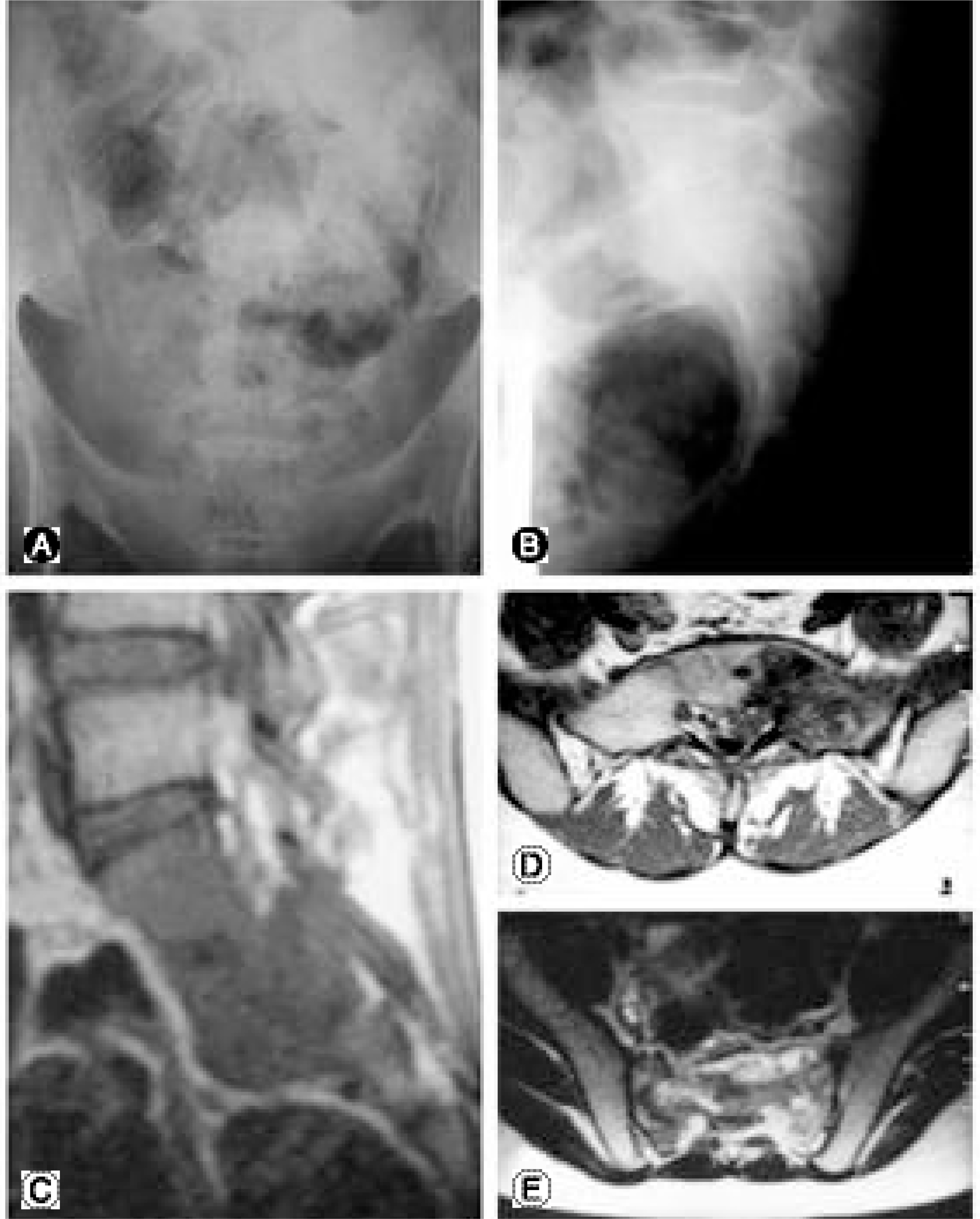
Fig. 1. Preoperative radiographic studies of case 1. AP (A) and lateral (B) plain roentgenographs show radiopacity at left sided upper half of sacrum. Sagittal MRI (C) shows the involvement of tumor at mid sacrum and axial MRI (D, E) show that the tumor involves especially in left side of sacrum.
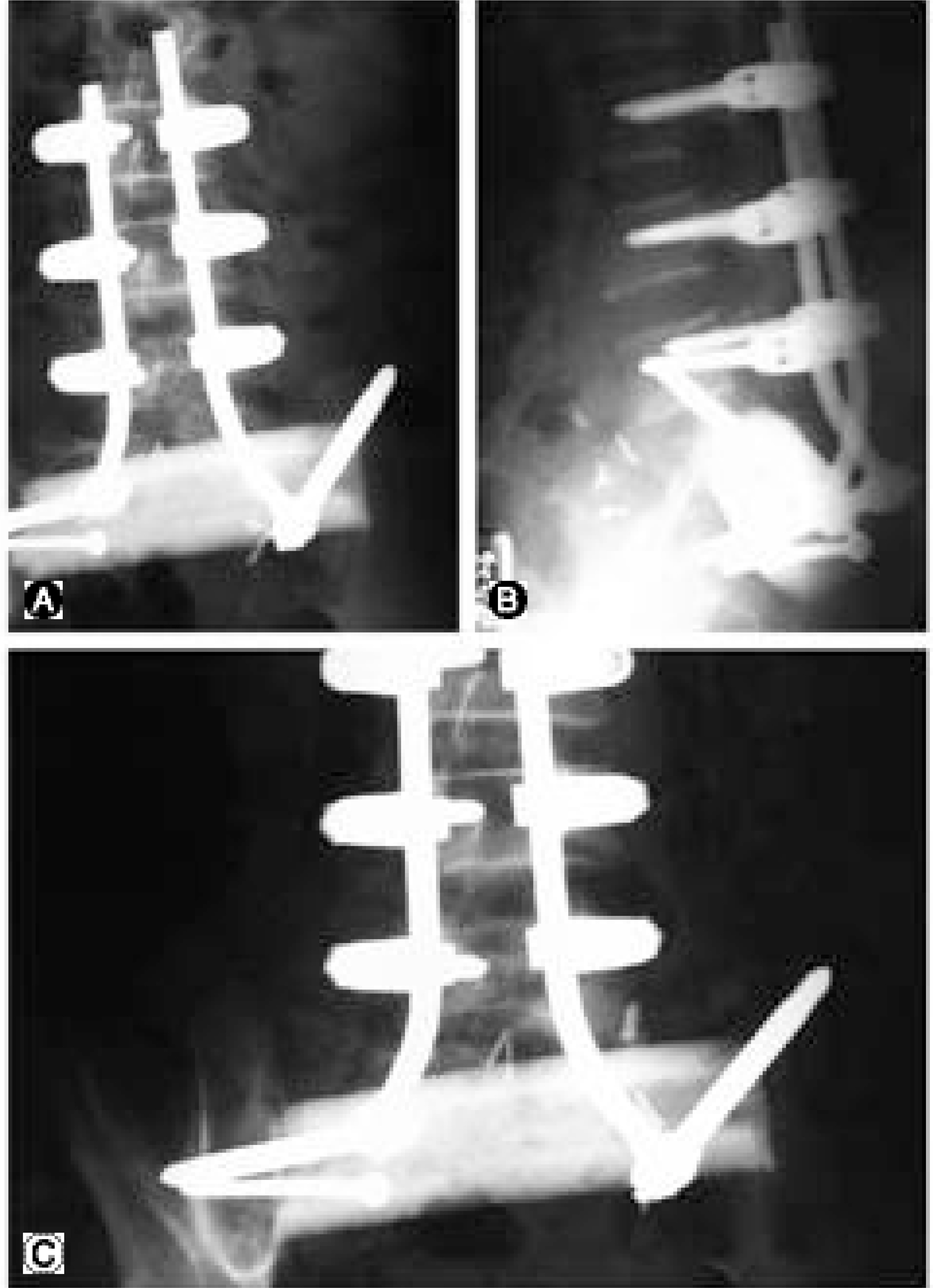
Fig. 2. Postoperative AP (A) and lateral (B) features of case 1. Sacrum is totally excised including both sacroiliac joints. The strut allograft is inserted thorough to both iliac wings and the instrumented reconstruction is done. At latest follow-up, union of grafted site and the maintenance of construct is seen (C).
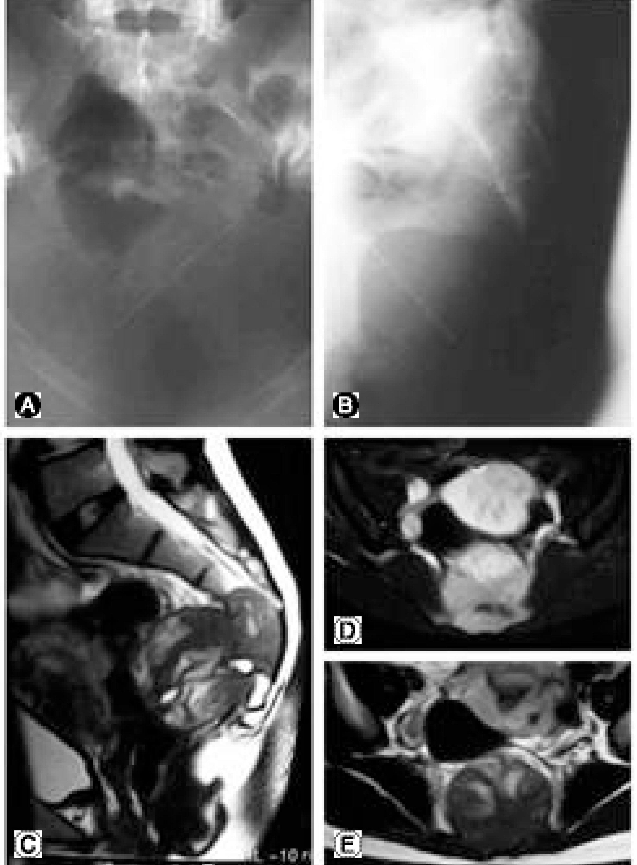
Fig. 3. Preoperative radiographic studies of case 2. AP (A) and lateral (B) plain roentgenographs show radiolucency at the lower half of sacrum. Sagittal T-2 weighted MRI (C), aixal T-1 weighted MRI (D), and axial T-2 weighted MRI (E) show the that the tumor mass is arising from lower sacrum and expending into the pelvic cavity.
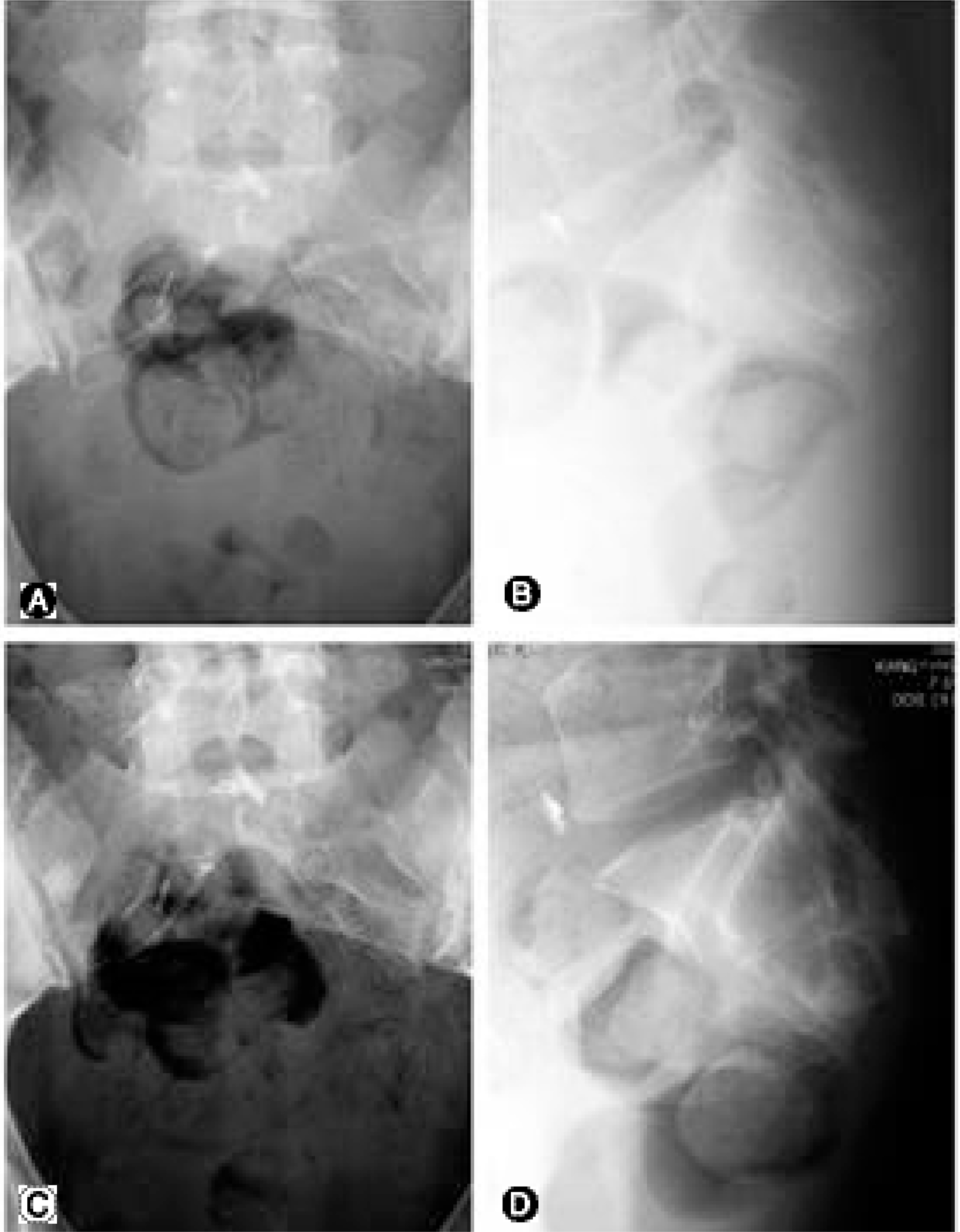
Fig. 4. Postoperative AP (A) and lateral (B) features of case 2. Sacrum and coccyx are totally excised from S3 level. At latest follow-up AP (C) and lateral (D) films show no definite change at operative site.
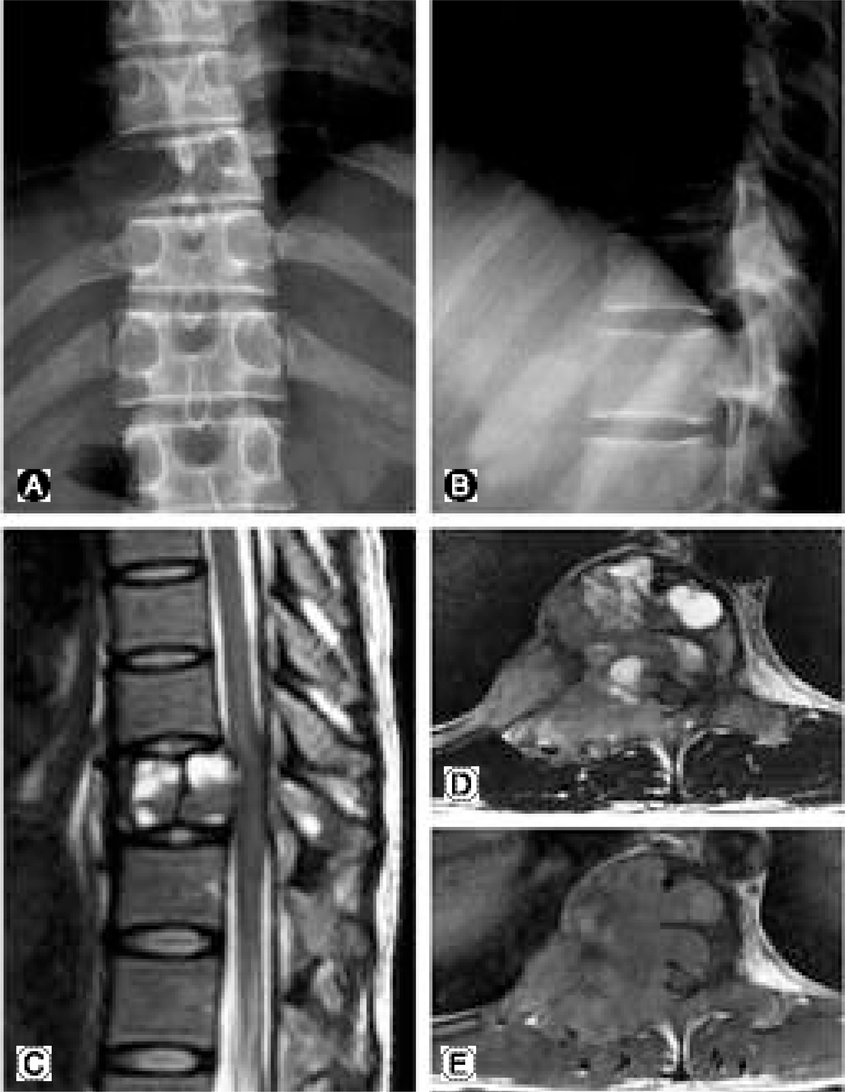
Fig. 5. Preoperative radiographic studies of case 3. AP (A) and lateral (B) plain roentgenographs show radiolucency at T10 vertebra including right pedicle. Sagittal T-2 weighted MRI (C), aixal T-2 weighted MRI (D), and axial T-1 weighted MRI (E) show the that the tumor mass involves entire T10 vertebral body, right sided pedicle, and the proximal portion of right sided 10th rib. The mass is protruding into the spinal canal and the spinal cord is compressed by tumor mass.
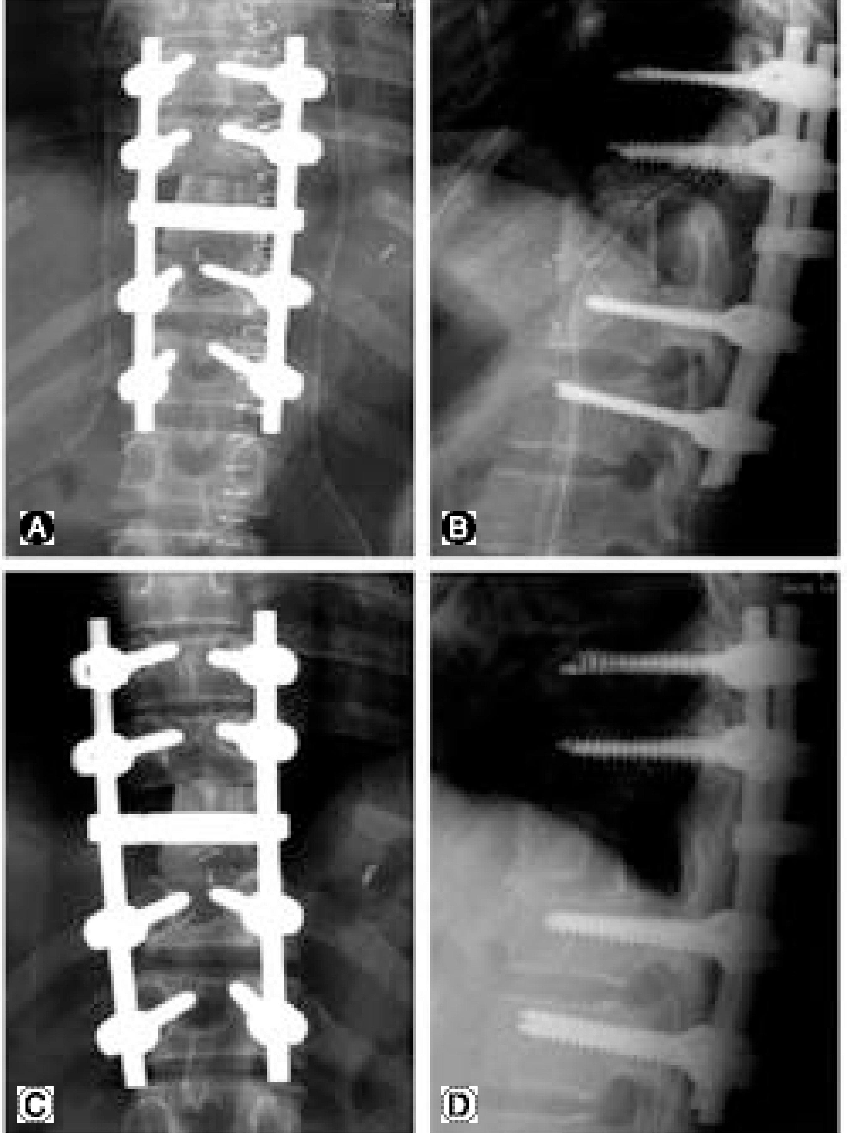
Fig. 6. Postoperative AP (A) and lateral (B) features of case 3. T10 vertebra and tumor mass are totally excised including proximal portion of both sided 10th ribs. At latest AP (C) and lateral (D) films show the union of lower side of allograft. At upper portion of allograft. there is no definite union but the evidence of healing can be found. The total feature of reconstruction is well maintained.
Reference
-
1). Abe E, Kobayashi T, Murai H, Suzuki T, Chiba M, Okuyama K. Total spondylectomy for primary malignant, aggressive benign, and solitary metastatic bone tumors of the thoracolumbar spine. J Spinal Disord. 2001; 14:237–246.
Article2). Doita M, Marui T, Nishida K, Kurosaka M, Yoshiya S, Sha N. Anterior spinal artery syndrome after total spondylectomy of T10, T11, and T12. Clin Orthop. 2002; 405:175–181.
Article3). Fidler MW. Surgical treatment of giant cell tumors of the thoracic and lumbar spine: report of nine patients. Eur Spine J. 2001; 10:69–77.4). Rhyu KW, Kang YK, Kwon SY, et al. Giant cell tumor of the lumbar spine treated with anterior lumbar interbody fusion using allograft and anterior and posterior combined fixation - a case report. J Korean Musculo Tissue Transp Soc. 2001; 1:149–155.5). Sakaura H, Hosono N, Mukai Y, Ishii T, Yonenobu K, Yoshikawa H. Outcome of total en bloc spondylectomy for solitary metastasis of the thoracolumbar spine. J Spinal Disord Tech. 2004; 17:297–300.
Article6). Stener B, Johnson OE. Complete removal of three vertebrae for giant-cell tumor. J Bone Joint Surg. 1971; 53-B:278–287.7). Tomita K, Kawahara N, Baba H, Tsuchiya H, Fujita T, Toribadake Y. Total en bloc spondylectomy: a new surgical techniques for primary malignant vertebral tumors. Spine. 1997; 22:324–333.8). Tomita K, Toribadake Y, Kawahara N, Ohnari H, Kose H. Total en bloc spondylectomy and circumspinal decompression for solitary spinal metastasis. Paraplegia. 1994; 32:36–46.
Article9). van Dijk M, Cuesta MA, Wuisman PIJM. Th oraco -scopically assisted total en bloc spondylectomy. Surg Endoc. 2000; 14:849–852.10). Anand N, Regan JJ. Video-assisted thoracoscopic surgery for the thoracic disc disease: classification and outcome study of 100 consecutive cases with a 2-year min -imun follow-up period. Spine. 2002; 27:871–879.11). Arlet V. Anterior thoracoscopic spine release in deformity surgery: a metaanalysis and review. Eur Spine J. 2000; 9(Suppl 1):S17–S23.
Article12). Kulko TR, Lenke LG. Thoracoscopic spine surgery: current indications and techniques. Orthop Nurs. 2000; 19:15–22.13). Lenke LG. Anterior endoscopic discectomy and fusion for adolescent idiopathic scoliosis. Spine. 2003; 28(Suppl 15):S36–S43.
Article14). Mack MJ, Regan JJ, McAfee PC, Picetti G, Ben-Yishay A, Acuff TE. Video-assisted thoracic surgery for the anterior approach to the thoracic spine. Ann Thoracic Surg. 1995; 59:1100–1106.
Article15). Mathews HH, Long BH. Minimally invasive techniques for the treatment of intervertebral disk herniation. J Am Acad Orthop Surg. 2002; 10:80–85.
Article16). Picetti GD III, Ertl JP, Bueff HU. Endoscopic instrumentation, correction, and fusion of idiopathic scoliosis. Spine J. 2001; 1:190–197.17). Regan JJ, Aronoff RJ, Ohnmeiss DD, Sengupta DK. Laparoscopic approach to L4-5 for interbody fusion using BAK cages: Experience in the first 58 cases. Spine. 1999; 24:2171–2174.18). Rosenthal D. Endoscopic approaches to the thoracic spine. Eur Spine J. 2000; 9(Suppl 1):S8–S16.
Article19). Zdeblick TA. Laparoscopic spinal fusion. Orthop Clin North Am. 1998; 29:635–645.
Article20). Zucherman JF, Zdeblick TA, Bailey SA, Mahvi D, Hsu KY, Kohrs D. Instrumented laparoscopic spinal fusion: Preliminary results. Spine. 1995; 20:2029–2035.21). Buttermann GR, Glazer PA, Bradford DS. The use of bone allografts in the spine. Clin Orthop. 1996; 324:75–85.
Article22). Cohen DB, Chotivichit A, Fujita T, et al. Pseudoarthrosis repair. autogenous iliac crest versus femoral ring allograft. Clin Orthop. 2000; 371:46–55.23). Wimmer C, Krismer M, Gluch H, Ogon M, Stockl B. Autogenic versus allogenic bone graft in anterior lumbar interbody fusion. Clin Orthop. 1999; 360:122–126.
- Full Text Links
-
- Actions
-
Cited
- CITED
-
- Close
- Share
-
- Similar articles
-
- Thoracoscopic Anterior Release of the Spine in Total en Bloc Spondylectomy for Primary Thoracic Spinal Tumor : A case report
- Total Spondylectomy of a Lumber Vertebra with Giant Cell Tumor: One Case Report
- Combined en Bloc Spondylectomy and Chest Wall Resection for Malignant Tumors Invading Spinal Column and Chest Wall
- Total Body Replacement with an Expandable Cage after en Bloc Lumbar Spondylectomy
- Total Spondylectomy for Giant Cell Tumor of Cervical Spine
