Preoperative Embolization of Cerebellar Hemangioblastoma with Onyx: Report of Three Cases
- Affiliations
-
- 1Department of Diagnostic Radiology, Busan Paik Hospital, Inje University, Busan, Korea. hwjeong2000@lycos.co.kr
- 2Department of Neurology, Busan Paik Hospital, Inje University, Busan, Korea.
- 3Department of Neurosurgery, Busan Paik Hospital, Inje University, Busan, Korea.
- KMID: 1910774
- DOI: http://doi.org/10.5469/neuroint.2014.9.1.45
Abstract
- Hemangioblastoma is a benign and highly vascular tumor. Complete surgical resection of highly vascular tumor such as hemangioblastoma may be challenging due to excessive bleeding. Preoperative embolization of these lesions may decrease the intraoperative blood loss and facilitate excision. We report three cases of cerebellar hemangioblastomas that were embolized using Onyx.
Figure
-
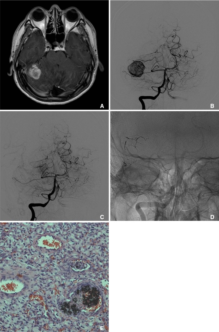
Fig. 1 A. Brain MRI (Contrast-enhanced T1-weighted image) reveals a 2.7 × 2.5 cm sized heterogeneously enhancing tumor with multiple signal void vascular structure within the tumor in the right cerebellar hemisphere. B. Diagnostic angiogram reveals the high vascularity with AV shunting within the tumor, which is characteristic for hemangioblastoma. C. The patient undergoes embolization of two distal pedicles off the right superior cerebellar artery using Onyx, resulting in near complete resolution of the tumor blush. D. Native cranial view in posteroanterior projection demonstrates a dense Onyx cast within mural nodule of the tumor. E. Histological examination of the excised tumor shows that the hemangioblastoma is composed of stromal cells, which show characteristically large and vacuolated appearance, and abundant vascular cells, such as endothelial cells and pericytes. Nidus vessels are occluded with thrombus and black colored embolic material, Onyx.

Fig. 2 A. Brain MRI (Contrast-enhanced T1-weighted image) reveals a cystic tumor with a densely enhancing mural nodule in the right cerebellum. B. A diagnostic vertebral angiogram reveals early intense tumor staining (arrows) in the right cerebellum from the right superior cerebellar artery. C. The patient undergoes embolization of core feeder vessel originated off the right superior cerebellar artery using Onyx. Final angiogram shows near complete resolution of the tumor blush.

Fig. 3 A. Brain MRI (Contrast-enhanced T1-weighted image) reveals a large cystic tumor with a heterogeneously enhancing mural nodule in the right cerebellum with tumor effect causing displacement of the fourth ventricle. B. A diagnostic vertebral angiogram reveals early intense tumor staining in the right cerebellum from the right superior cerebellar artery. C. The patient undergoes embolization of core feeder vessel originated off the right superior cerebellar artery using Onyx. Final angiogram shows near complete resolution of the tumor blush.
Reference
-
1. Horvathy DB, Hauch EF, Ogilvy CS, Hopkins LN, Levy EI, Siddiqui AH. Complete preoperative embolization of hemangioblastoma vessels with Onyx 18. J Clin Neurosci. 2011; 18:401–403. PMID: 21237650.
Article2. Dabus G, Pryor J, Spilberg G, Samaniego EA, Nogueira RG. Embolization of intra-axial hypervascular tumors with Onyx: report of three cases. J Neurointerv Surg. 2013; 5:177–180. PMID: 22266792.
Article3. Gore P, Theodore N, Brasiliense L, Kim LJ, Garrett M, Nakaji P, et al. The utility of Onyx for preoperative embolization of cranial and spinal tumor. Neurosurgery. 2008; 62:1204–1212. PMID: 18824987.4. Takeuchi S, Tanaka R, Fujii Y, Abe H, Ito Y. Surgical treatment of hemangioblastomas with presurgical endovascular embolization. Neurol Med Chir (Tokyo). 2001; 41:246–251. PMID: 11396304.
Article5. Eskridge JM, McAuliffe W, Harris B, Kim DK, Scott J, Winn HR. Preoperative endovascular embolization of craniospinal hemanigoblastoma. AJNR Am J Neuroradiol. 1996; 17:525–531. PMID: 8881249.6. Cornelius JF, Saint-Maurice JP, Bresson D, George B, Houdart E. Hemorrhage after particle embolization of hemangioblastomas: comparison of outcomes in spinal and cerebellar lesions. J Neurosurg. 2007; 106:994–998. PMID: 17564170.
Article7. Seong Eom K, Won Kim D, Sung Choi S, Ha Choi K, Young Kim T. Preoperative embolization of a cerebellar hemangioblastoma using Onyx. Neurol Neurochir Pol. 2011; 45(3):292–296. PMID: 21866486.8. Gobin YP, Murayama Y, Milanese K, Chow K, Gonzalez NR, Duckwiler GR, et al. Head and neck hypervascular lesions: embolization with ethylene vinyl alcohol copolymer. Radiology. 2001; 221:309–317. PMID: 11687669.
- Full Text Links
-
- Actions
-
Cited
- CITED
-
- Close
- Share
-
- Similar articles
-
- Cerebellar Hemangioblastoma:Hemorrhage as an Initial Presentation: Case Report
- A Case Report of Recurrent and Disseminated Cerebellar Hemangioblastoma
- Preoperative Embolization of Extra-axial Hypervascular Tumors with Onyx
- Complication Associated with Onyx Embolization of Spinal Cord Arteriovenous Malformation
- Hairball-Like Migration of “Onyx Threads” into the Draining Vein during Transarterial Embolization of a Dural Arteriovenous Fistula: A Case Report and Experimental Validation
