Preoperative Embolization of Extra-axial Hypervascular Tumors with Onyx
- Affiliations
-
- 1Department of Neurological Surgery, Vanderbilt University Medical Center, Nashville, TN, USA.
- 2Division of Neurosurgery, Department of Surgery, Beth Israel Deaconess Medical Center and Harvard Medical School, Boston, MA, USA.
- 3Department of Neurosurgery, Barrow Neurological Institute, Phoenix, AZ, USA. Bgross83@gmail.com
- KMID: 2161697
- DOI: http://doi.org/10.7461/jcen.2016.18.1.12
Abstract
OBJECTIVE
Preoperative endovascular embolization of intracranial tumors is performed to mitigate anticipated intraoperative blood loss. Although the usage of a wide array of embolic agents, particularly polyvinyl alcohol (PVA), has been described for a variety of tumors, literature detailing the efficacy, safety and complication rates for the usage of Onyx is relatively sparse.
MATERIALS AND METHODS
We reviewed our single institutional experience with pre-surgical Onyx embolization of extra-axial tumors to evaluate its efficacy and safety and highlight nuances of individualized cases.
RESULTS
Five patients underwent pre-surgical Onyx embolization of large or giant extra-axial tumors within 24 hours of surgical resection. Four patients harbored falcine or convexity meningiomas (grade I in 2 patients, grade II in 1 patient and grade III in one patient), and one patient had a grade II hemangiopericytoma. Embolization proceeded uneventfully in all cases and there were no complications.
CONCLUSION
This series augments the expanding literature confirming the safety and efficacy of Onyx in the preoperative embolization of extra-axial tumors, underscoring its advantage of being able to attain extensive devascularization via only one supplying pedicle.
Keyword
Figure
-

Fig. 1 Case 1. Preoperative T1-weighted post contrast coronal magnetic resonance imaging showing an extra-axial mass with significant mass effect and edema on the adjacent brain parenchyma (A). Anteroposterior view of left external carotid artery arteriogram demonstrating filling of the tumor through branches of the left superficial temporal artery and the left middle meningeal artery draining into a venous fistula and also supplying the tumor through multiple collaterals (B). Anteroposterior skull film demonstrating Onyx cast (C).

Fig. 2 Case 2. Preoperative T1-weighted post contrast coronal magnetic resonance imaging demonstrates an extra-axial, falcine lesion over the frontoparietal vertex with mass effect and an associated inferolateral peritumoral cyst (A). Anteroposterior view of a distal left external carotid artery arteriogram illustrates the classic "spoke wheel" filling of the falcine meningioma supplied by distal branch of the middle meningeal artery (C). Lateral view of post-embolization angiogram of left external carotid artery shows occlusion of the distal middle meningeal artery.
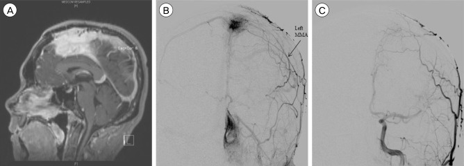
Fig. 3 Case 3. Preoperative T1-weighted post contrast sagittal magnetic resonance imaging shows an extra-axial enhancing mass centered in the falx cerebri (A). This lesion extended bilaterally with invasion of the anterior and mid superior sagittal sinus. Anteroposterior view of left external carotid artery arteriogram showing multiple arterial feeders to the tumor, mainly from the left middle meningeal artery (B). Anteroposterior view of post-embolization left common carotid artery arteriogram (C).
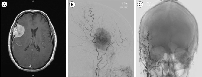
Fig. 4 Case 4. Preoperative T1-weighted post contrast axial magnetic resonance imaging shows a right frontal extra-axial mass with intraosseous extension and adjacent dural thickening (A). Lateral view of right external carotid artery angiogram shows a hypervascular tumor blush, supplied by the middle meningeal artery and accessory middle meningeal artery (B). Anteroposterior, unsubtracted view of post-embolization of right external carotid artery angiogram demonstrates Onyx casting within the distal middle meningeal artery, with markedly diminished supply to the tumor but little flow from the accessory middle meningeal artery (C).

Fig. 5 Case 5. Preoperative T1-weighted post contrast coronal magnetic resonance imaging shows a right temporal occipital extra-axial mass, with midline shift and mass effect noted on the right lateral ventricle and extensive edema (A). Anteroposterior view of right external carotid artery angiogram shows arterial supply to the tumor from branches of right occipital artery and the middle meningeal artery (B). Lateral view of post-embolization of right common carotid artery angiogram demonstrates little residual supply to the tumor (C).
Reference
-
1. Ding D, Kreitel D, Liu KC. Onyx embolization of an intracranial hemangiopericytoma by direct transcranial puncture. Interv Neuroradiol. 2013; 12. 19(4):466–470. PMID: 24355151.
Article2. Dowd CF, Halbach VV, Higashida RT. Meningiomas: the role of preoperative angiography and embolization. Neurosurg Focus. 2003; 7. 15(1):E10. PMID: 15355012.
Article3. Elhammady MS, Peterson EC, Johnson JN, Aziz-Sultan MA. Preoperative onyx embolization of vascular head and neck tumors by direct puncture. World Neurosurg. 2012; May-Jun. 77(5-6):725–730. PMID: 22079824.
Article4. Gobin YP, Murayama Y, Milanese K, Chow K, Gonzalez NR, Duckwiler GR, et al. Head and neck hypervascular lesions: embolization with ethylene vinyl alcohol copolymer--laboratory evaluation in Swine and clinical evaluation in humans. Radiology. 2001; 11. 221(2):309–317. PMID: 11687669.
Article5. Gore P, Theodore N, Brasiliense L, Kim LJ, Garrett M, Nakaji P, et al. The utility of onyx for preoperative embolization of cranial and spinal tumors. Neurosurgery. 2008; 6. 62(6):1204–1211. PMID: 18824987.
Article6. Kai Y, Hamada J, Morioka M, Yano S, Todaka T, Ushio Y. Appropriate interval between embolization and surgery in patients with meningioma. AJNR Am J Neuroradiol. 2002; 1. 23(1):139–142. PMID: 11827886.7. Pierot L, Cognard C, Herbreteau D, Fransen H, van Rooi WJ, Boccardi E, et al. Endovascualr treatment of brain arteriovenous malformations using a liquid emboilc agent: results of a prospective, multicentre study (BRAVO). Eur Radiol. 2013; 10. 23(10):2838–2845. PMID: 23652849.8. Rangel-Castilla L, Shah AH, Klucznik RP, Diaz OM. Preoperative Onyx embolization of hypervascular head, neck, and spinal tumors: experience with 100 consecutive cases from a single tertiary center. J Neurointerv Surg. 2014; 1. 6(1):51–56. PMID: 23268473.
Article9. Rossitti S. Preoperative embolization of lower-falx meningiomas with ethylene vinyl alcohol copolymer: technical and anatomical aspects. Acta Radiol. 2007; 4. 48(3):321–326. PMID: 17453504.
Article10. Shah AH, Patel N, Raper DM, Bregy A, Ashour R, Elhammady MS, et al. The role of preoperative embolization for intracranial meningiomas. J Neurosurg. 2013; 8. 119(2):364–372. PMID: 23581584.
Article11. Shi ZS, Feng L, Jiang XB, Huang Q, Yang Z, Huang ZS. Therapeutic embolization of meningiomas with Onyx for delayed surgical resection. Surg Neurol. 2008; 11. 70(5):478–481. PMID: 18261767.
Article12. Taki W, Yonekawa Y, Iwata H, Uno A, Yamashita K, Amemiya H. A new liquid material for embolization of arteriovenous malformations. AJNR AM J Neuroradiol. 1990; Jan-Feb. 11(1):163–168. PMID: 2105599.
- Full Text Links
-
- Actions
-
Cited
- CITED
-
- Close
- Share
-
- Similar articles
-
- Preoperative Embolization of Hypervascular Brain Tumor Fed by Branches of the Internal Carotid Artery
- Complication Associated with Onyx Embolization of Spinal Cord Arteriovenous Malformation
- Preoperative Embolization of Cerebellar Hemangioblastoma with Onyx: Report of Three Cases
- Hairball-Like Migration of “Onyx Threads” into the Draining Vein during Transarterial Embolization of a Dural Arteriovenous Fistula: A Case Report and Experimental Validation
- Therapeutic Embolization of Renal Tumor
