Extravasation of Joint Fluid into the Mediastinum and the Deep Neck during Arthroscopic Shoulder Surgery
- Affiliations
-
- 1Department of Radiology, Dong-A University Hospital, Dong-A University College of Medicine, Busan, Korea. gnlee@dau.ac.kr
- KMID: 1897259
- DOI: http://doi.org/10.3348/jksr.2014.70.1.43
Abstract
- Extravasation of shoulder joint fluid into the surrounding muscles during shoulder arthroscopic surgery is common and inevitable. Here, we report a case of massive extravasation of shoulder joint fluid leading to mediastinal and retrotracheal effusion after arthroscopic shoulder surgery. We will discuss the anatomical basis of fluid leakage from the shoulder to the mediastinum and to the deep neck on CT.
Figure
-
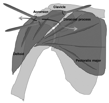
Fig. 1 Spaces of potential fluid leakage from the shoulder. Fluid extravasation occurs when arthroscopic shoulder surgery is performed at the subacromial space. Three ports were inserted near the acromion [anterolateral, posterior (not seen in the figure), and lateral to the acromion], and the anteroinferior port was inserted near the coracoid process. Spaces of potential fluid leakage are along the margin of the deltoid muscle and through the arthroscopic port that penetrates the deltoid muscle (arrows). A large amount of fluid might have accumulated in the subcutaneous fat anterior to the pectoralis major muscle in this course.

Fig. 2 Fluid leakage during shoulder arthroscopy in a 59-year-old woman. A. Enhanced chest CT scan shows a large amount of fluid accumulation in the subcutaneous fat layer of the anterior chest wall (star). Fluid is also noted in the posterior chest wall, permeating the right rhomboid and the right latissimusdorsi muscles (arrows). B. Enhanced chest CT scan at the level of the upper neck shows fluid in the retropharyngeal space (star), right posterior cervical space (triangle), and right submandibular space (square). Fluid splayed between the carotid space and anterior scalene muscle to enter into the retropharyngeal space of the neck (arrow). C. Thin-section CT scan of the lung shows smooth interlobular septal thickening in the right apex and the focal ground glass opacity in left apex medial aspect (arrow). D. Coronal-reformatted chest CT scan shows that the fluid around the subclavian vein continues to the mediastinum (arrows).
Reference
-
1. Berjano P, González BG, Olmedo JF, Perez-España LA, Munilla MG. Complications in arthroscopic shoulder surgery. Arthroscopy. 1998; 14:785–788.2. Hynson JM, Tung A, Guevara JE, Katz JA, Glick JM, Shapiro WA. Complete airway obstruction during arthroscopic shoulder surgery. Anesth Analg. 1993; 76:875–878.3. Borgeat A, Bird P, Ekatodramis G, Dumont C. Tracheal compression caused by periarticular fluid accumulation: a rare complication of shoulder surgery. J Shoulder Elbow Surg. 2000; 9:443–445.4. Blumenthal S, Nadig M, Gerber C, Borgeat A. Severe airway obstruction during arthroscopic shoulder surgery. Anesthesiology. 2003; 99:1455–1456.5. Orebaugh SL. Life-threatening airway edema resulting from prolonged shoulder arthroscopy. Anesthesiology. 2003; 99:1456–1458.6. Antonucci S, Orlandi P, Mattei PA, Amato F. Airway obstruction during arthroscopic shoulder surgery: anesthesia for the patient or for the surgeon? Minerva Anestesiol. 2006; 72:995–1000.7. Jirativanont T, Tritrakarn TD. Upper airway obstruction following arthroscopic rotator cuff repair due to excess irrigation fluid. Anaesth Intensive Care. 2010; 38:957–958.8. Ogilvie-Harris DJ, Boynton E. Arthroscopic acromioplasty: extravasation of fluid into the deltoid muscle. Arthroscopy. 1990; 6:52–54.9. Harnsberger HR. Suprahyoid and infrahyoid neck. In : Harnsberger HR, Osborn AG, MacDonald AJ, Ross JS, Moore KR, Salzman KL, editors. Diagnostic and surgical imaging anatomy: Brain, Head & Neck, Spine. Salt Lake City, UT: Amirsys publishing Inc;2011. p. II126–II252.10. Jeong YJ, Kim S, Kwak SW, Lee NK, Lee JW, Kim KI, et al. Neoplastic and nonneoplastic conditions of serosal membrane origin: CT findings. Radiographics. 2008; 28:801–817.
- Full Text Links
-
- Actions
-
Cited
- CITED
-
- Close
- Share
-
- Similar articles
-
- Pleural effusion caused by extravasation of irrigation fluid during arthroscopic shoulder surgery: A case report
- Life-threatening Airway Edema after Arthroscopic Repair of Massive Rotator Cuff Tear: A Case Report
- Fatal Pulmonary Edema During Arthroscopic Shoulder Surgery
- Does desflurane need more irrigating-pump pressure for the visibility in arthroscopic shoulder surgery than sevoflurane?
- Current Concepts in Arthroscopic Treatment of Anterior Shoulder Instability
