Clinical Features and Treatments of Carpal Bone Cysts
- Affiliations
-
- 1Department of Orthopedic Surgery, Sun General Hospital, Daejeon, Korea. skrhee@catholic.ac.kr
- 2Department of Orthopedic Surgery, Yeouido St. Mary's Hospital, The Catholic University of Korea College of Medicine, Seoul, Korea.
- KMID: 1896921
- DOI: http://doi.org/10.12790/jkssh.2014.19.1.7
Abstract
- PURPOSE
A total of 27 carpal bone cysts were analyzed for their sites, relations of other wrist soft tissue ganglions and their results of treatment were evaluated.
METHODS
Twenty-seven carpal bone cysts in 20 patients (bilateral 5, multiple 2) from February 2002 to June 2013 were evaluated. Mean follow-up period was 16.6 months. We investigated etiological classification, the site of carpal bone cyst, and their relationship with soft tissue ganglion in same wrist. Pain, range of motion, radiographic changes, and their satisfaction after treatment were assessed postoperatively.
RESULTS
The carpal bone cysts occurred mainly at the radial wrist axial ray on the lunate (12 cases), scaphoid (6 cases), and triquetrum (5 cases), trapezium (2 cases), and capitate (2 cases). Based on the magnetic resonance imaging (MRI) findings in 25 cases, we classified carpal bone cysts into 4 distinct types; type I with purely intraosseous lesion (16 cases), type II with bone cyst associated cortical perforations (6 cases), type III with coexisting soft tissue ganglion communicating with intra-osseous lesion (2 cases), and type IV with coexisting soft tissue ganglion non-communicating intraosseous lesions (1 case).
CONCLUSION
The carpal bone cysts can be classified by MRI into 4 distinct types. The purely intraosseous type is most common, suggesting the intrinsic cause in the development of carpal bone cyst.
Keyword
MeSH Terms
Figure
-
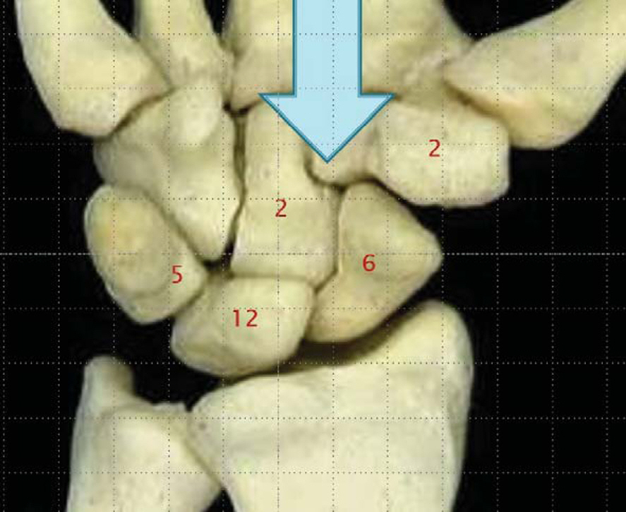
Fig. 1. The location of carpal bone cysts.
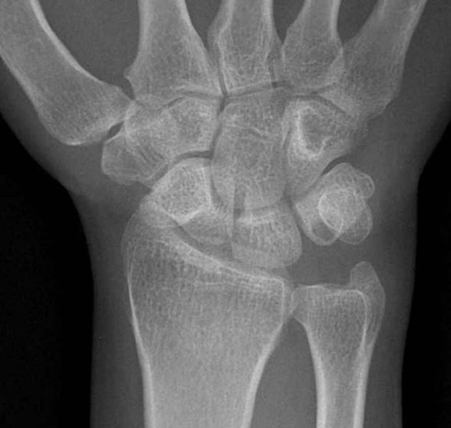
Fig. 2. Anteroposterior radiograph of wrist shows radiolucent lesion on triquetrum, purely intraosseous bone cyst.
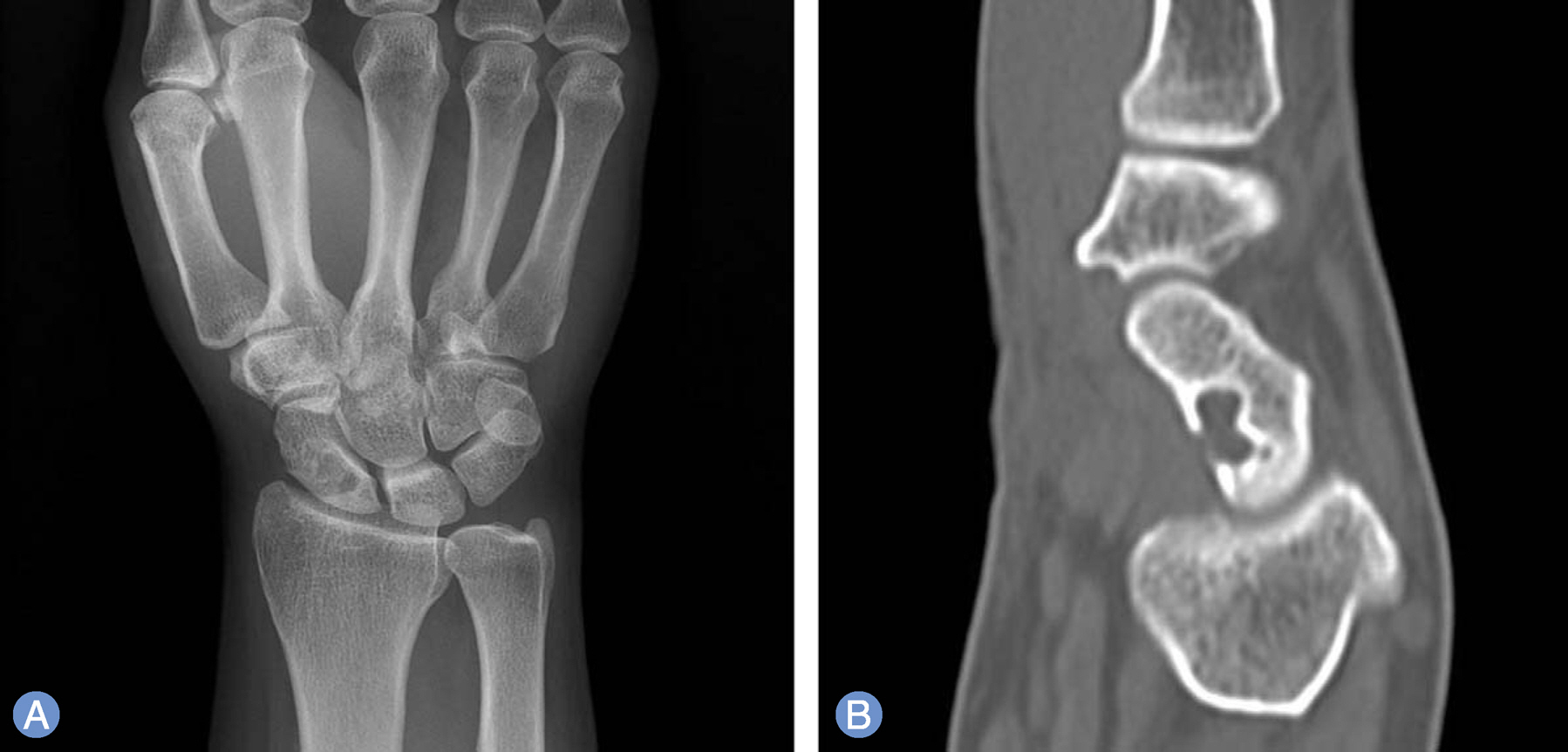
Fig. 3. (A) Anteroposterior radiograph of wrist shows radiolucent lesion on scaphoid. (B) Saggital computed tomography showing the lytic intraosseous ganglion on scaphoid with a sclerotic margin, expansion and perforating of the cortex.
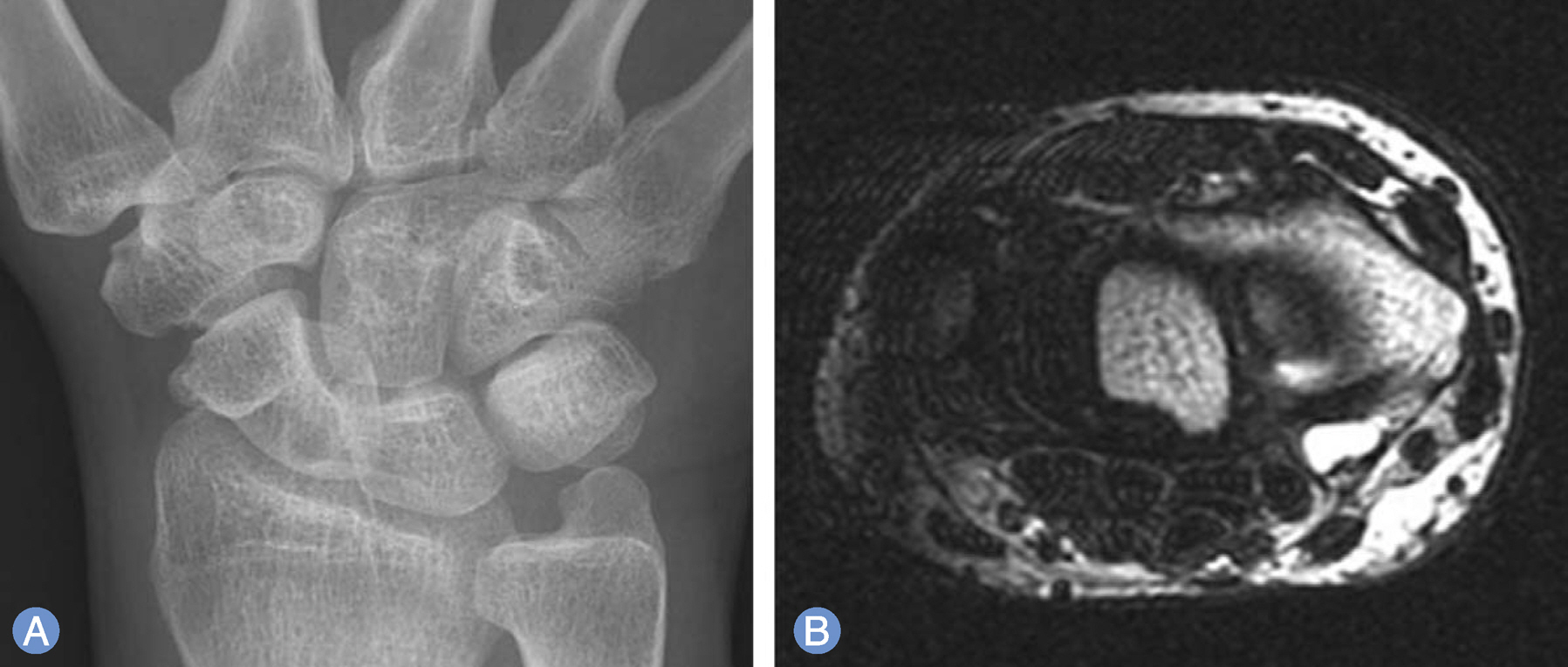
Fig. 4. (A) Anteroposterior radiograph of wrist shows radiolucent lesion on scaphoid. (B) Axial T1-weighted magnetic resonance imaging shows slightly hypointensity signal change close to the volar soft tissue ganglion.
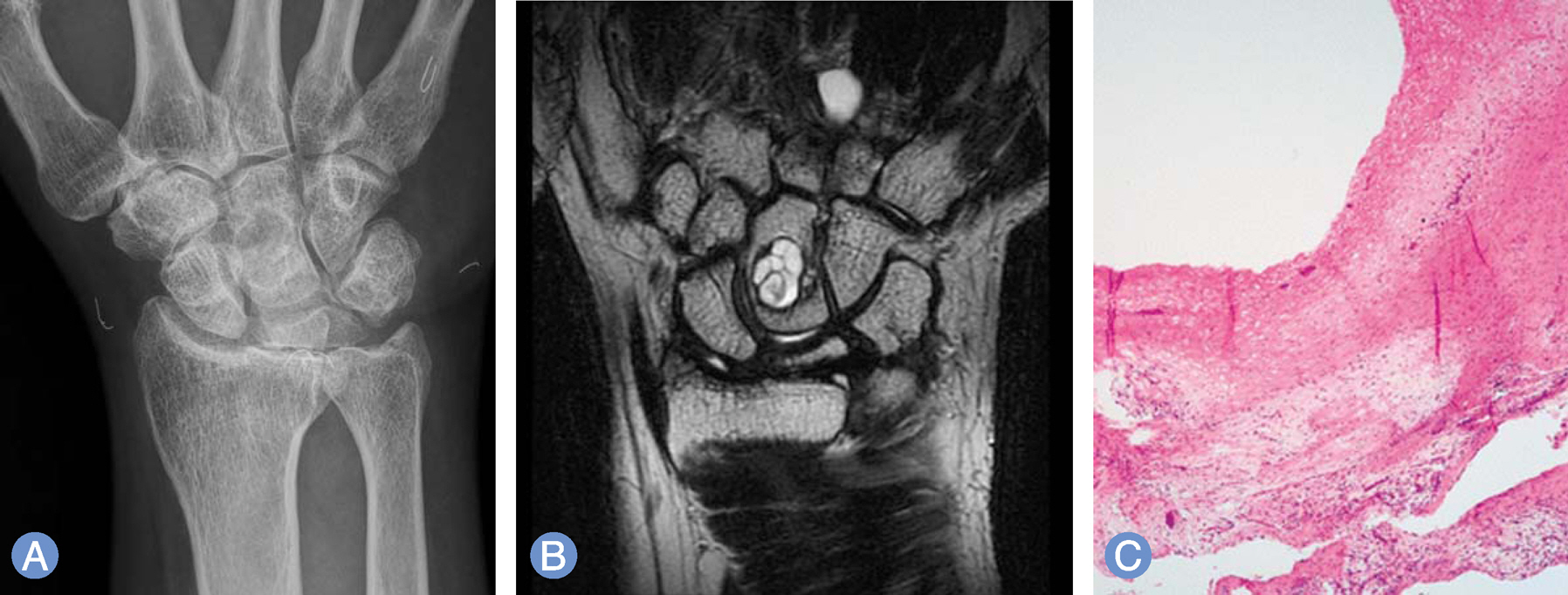
Fig. 5. (A) Anteroposterior radiograph of wrist shows radiolucent lesion on capitate. (B) Coronal T1-weighted magnetic resonance imaging shows tissue ganglion non-communicating with intra-osseous bone cyst on capitate. (C) The histology shows a multiloculated, cystic lesion with foci of myxoid degeneration in the surrounding stroma (H&E, ×40).
Reference
-
References
1. Schajowicz F, Clavel Sainz M, Slullitel JA. Juxta-articular bone cysts (intra-osseous ganglia): a clinicopathological study of eighty-eight cases. J Bone Joint Surg Br. 1979; 61:107–16.
Article2. Uriburu IJ, Levy VD. Intraosseous ganglia of the scaphoid and lunate bones: report of 15 cases in 13 patients. J Hand Surg Am. 1999; 24:508–15.
Article3. Lorente R, Moreno M, Quiles M. Bilateral intraosseous ganglia of the lunate: a case report. J Hand Surg Am. 1992; 17:1084–5.
Article4. Logan SE, Gilula LA, Kyriakos M. Bilateral scaphoid ganglion cysts in an adolescent. J Hand Surg Am. 1992; 17:490–5.
Article5. Bowers WH, Hurst LC. An intraarticular-intraosseous carpal ganglion. J Hand Surg Am. 1979; 4:375–7.
Article6. Posner MA, Green SM. Intraosseous ganglion of a phalanx. J Hand Surg Am. 1984; 9:280–2.
Article7. Kambolis C, Bullough PG, Jaffe HI. Ganglionic cystic defects of bone. J Bone Joint Surg Am. 1973; 55:496–505.
Article8. Iwahara T, Hirayama T, Takemitu Y. Intraosseous ganglion of the lunate. Hand. 1983; 15:297–9.9. Suh JS, Lee SM, Kim DS. Intraosseous ganglion in capitate: a case report. J Korean Soc Surg Hand. 2003; 8:81–4.10. Han CW, Kim JC, Lee HK, Ha JD, Shin YH, Kim WY. Intraosseous ganglion of the lunate. J Korean Soc Surg Hand. 2003; 8:47–9.11. Park MR, Lee GH, Kim SH. Intraosseous ganglion in triquetrum: a case report. J Korean Orthop Assoc. 2000; 35:661–3.
Article12. Kligman M, Roffman M. Bilateral intraosseous ganglia of the scaphoid and lunate bones. J Hand Surg Br. 1997; 22:820–1.
Article13. Van den Dungen S, Marchesi S, Ezzedine R, Bindou D, Lorea P. Relationship between dorsal ganglion cysts of the wrist and intraosseous ganglion cysts of the carpal bones. Acta Orthop Belg. 2005; 71:535–9.14. Tham S, Ireland DC. Intraosseous ganglion cyst of the lunate: diagnosis and management. J Hand Surg Br. 1992; 17:429–32.
Article
- Full Text Links
-
- Actions
-
Cited
- CITED
-
- Close
- Share
-
- Similar articles
-
- Management of carpal bone fractures other than the scaphoid: a narrative review
- Intraosseous Ganglia of Scaphoid in Hand: Report of 2 Cases
- Ultrasound-Guided Nerve Hydrodissection for Carpal Tunnel Syndrome
- Incidence of Carpal Bone Injuries and It's Radiologic Consideration
- Essential Osteolysis of Carpal and Tarsal Bones: A Case Report
