Balloon Occlusive Diameter of Non-Circular Atrial Septal Defects in Transcatheter Closure with Amplatzer Septal Occluder
- Affiliations
-
- 1Department of Pediatrics, Samsung Medical Center, Sungkyunkwan University School of Medicine, Seoul, Korea. amyjys@naver.com
- 2Department of Medicine, Samsung Medical Center, Sungkyunkwan University School of Medicine, Seoul, Korea.
- KMID: 1826559
- DOI: http://doi.org/10.4070/kcj.2013.43.10.681
Abstract
- BACKGROUND AND OBJECTIVES
The aim of this study was to investigate the balloon occlusive diameter (BOD) of non-circular defects in the transcatheter closure of atrial septal defect (ASD).
SUBJECTS AND METHODS
A total of 67 patients who had undergone transcatheter closure of an ASD were reviewed retrospectively. A non-circular defect was defined as the ratio of the short diameter to the long diameter of the defect on the en-face image less than 0.75. The BOD was compared with the long diameter of the defect and then compared between the two groups.
RESULTS
There were 22 patients with circular defects and 45 patients with non-circular defects. The difference in BOD measuring from the long diameter of the defect was quite different between the two groups and significantly smaller in non-circular morphology (0.1+/-4.0 vs. 2.3+/-2.1, p=0.006). The difference in BOD measurement from the long diameter of ASD showed a positive correlation with the ratio of the short diameter to the long diameter of ASD (b/a) (r2=0.102, p=0.008). In the non-circular morphology of ASD, the difference in BOD measured from the long diameter had a significant negative correlation with the long diameter of ASD (r2=0.230, p=0.001), whereas in circular ASD, no significant correlation was found between the difference in BOD and the long diameter of ASD (p=0.201).
CONCLUSION
The BOD compared with the long diameter measured from three-dimensional transesophageal echocardiography was smaller in non-circular ASD than in circular ASD. This difference was much smaller in non-circular ASD with a large long diameter.
MeSH Terms
Figure
-
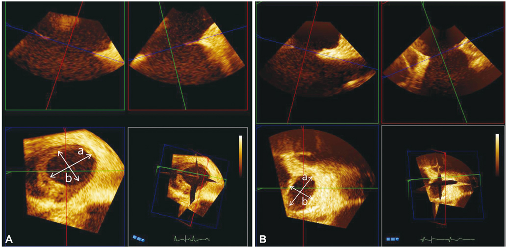
Fig. 1 The reconstructed en-face image from three-dimensional transesophageal echocardiography shows a circular defect (A) and a non-circular defect (B). a: the long diameter, b: the short diameter.
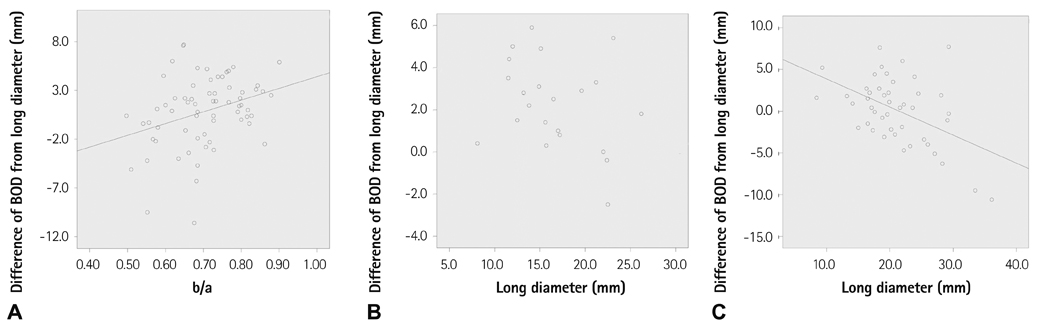
Fig. 2 The difference of the balloon occlusive diameter from the long diameter of the atrial septal defect (ASD) had a significant positive correlation with the ratio of the short diameter to the long diameter (A). The difference of balloon occlusive diameter from the defect size did not have a significant correlation with the long diameter in ASD with circular morphology (p=0.201) (B), whereas the difference in balloon occlusive diameter had a significant negative correlation with the long diameter in ASD with non-circular morphology (p=0.001) (C). BOD: balloon occlusive diameter.
Reference
-
1. Masura J, Gavora P, Podnar T. Long-term outcome of transcatheter secundum-type atrial septal defect closure using Amplatzer septal occluders. J Am Coll Cardiol. 2005; 45:505–507.2. Majunke N, Bialkowski J, Wilson N, et al. Closure of atrial septal defect with the Amplatzer septal occluder in adults. Am J Cardiol. 2009; 103:550–554.3. Yew G, Wilson NJ. Transcatheter atrial septal defect closure with the Amplatzer septal occluder: five-year follow-up. Catheter Cardiovasc Interv. 2005; 64:193–196.4. Kim NK, Park SJ, Choi JY. Transcatheter closure of atrial septal defect: does age matter? Korean Circ J. 2011; 41:633–638.5. Johri AM, Witzke C, Solis J, et al. Real-time three-dimensional transesophageal echocardiography in patients with secundum atrial septal defects: outcomes following transcatheter closure. J Am Soc Echocardiogr. 2011; 24:431–437.6. Roberson DA, Cui W, Patel D, et al. Three-dimensional transesophageal echocardiography of atrial septal defect: a qualitative and quantitative anatomic study. J Am Soc Echocardiogr. 2011; 24:600–610.7. Ferreira SM, Ho SY, Anderson RH. Morphological study of defects of the atrial septum within the oval fossa: implications for transcatheter closure of left-to-right shunt. Br Heart J. 1992; 67:316–320.8. van den Bosch AE, Ten Harkel DJ, McGhie JS, et al. Characterization of atrial septal defect assessed by real-time 3-dimensional echocardiography. J Am Soc Echocardiogr. 2006; 19:815–821.9. Bhaya M, Mutluer FO, Mahan E, et al. Live/Real time three-dimensional transesophageal echocardiography in percutaneous closure of atrial septal defects. Echocardiography. 2013; 30:345–353.10. Nasis A, Harper RW, Mottram PM. Real-time three-dimensional transoesophageal echocardiography for guidance of transcatheter closure of a complex multifenestrated atrial septal defect. Heart Lung Circ. 2011; 20:343–344.11. Marx GR, Fulton DR, Pandian NG, et al. Delineation of site, relative size and dynamic geometry of atrial septal defects by real-time three-dimensional echocardiography. J Am Coll Cardiol. 1995; 25:482–490.12. Huang X, Shen J, Huang Y, et al. En face view of atrial septal defect by two-dimensional transthoracic echocardiography: comparison to real-time three-dimensional transesophageal echocardiography. J Am Soc Echocardiogr. 2010; 23:714–721.13. Seo JS, Song JM, Kim YH, et al. Effect of atrial septal defect shape evaluated using three-dimensional transesophageal echocardiography on size measurements for percutaneous closure. J Am Soc Echocardiogr. 2012; 25:1031–1040.14. Chan KC, Godman MJ, Walsh K, Wilson N, Redington A, Gibbs JL. Transcatheter closure of atrial septal defect and interatrial communications with a new self expanding nitinol double disc device (Amplatzer septal occluder): multicentre UK experience. Heart. 1999; 82:300–306.15. Acar P. Three-dimensional echocardiography in transcatheter closure of atrial septal defects. Cardiol Young. 2000; 10:484–492.16. Ko SF, Liang CD, Yip HK, et al. Amplatzer septal occluder closure of atrial septal defect: evaluation of transthoracic echocardiography, cardiac CT, and transesophageal echocardiography. AJR Am J Roentgenol. 2009; 193:1522–1529.17. Zanchetta M. On-line intracardiac echocardiography alone for Amplatzer Septal Occluder selection and device deployment in adult patients with atrial septal defect. Int J Cardiol. 2004; 95:61–68.18. Amin Z, Hijazi ZM, Bass JL, Cheatham JP, Hellenbrand WE, Kleinman CS. Erosion of Amplatzer septal occluder device after closure of secundum atrial septal defects: review of registry of complications and recommendations to minimize future risk. Catheter Cardiovasc Interv. 2004; 63:496–502.19. Divekar A, Gaamangwe T, Shaikh N, Raabe M, Ducas J. Cardiac perforation after device closure of atrial septal defects with the Amplatzer septal occluder. J Am Coll Cardiol. 2005; 45:1213–1218.20. Sadeghian H, Hajizeinali A, Eslami B, et al. Measurement of Atrial Septal Defect Size: A Comparative Study between Transesophageal Echocardiography and Balloon Occlusive Diameter Method. J Tehran Heart Cent. 2010; 5:74–77.
- Full Text Links
-
- Actions
-
Cited
- CITED
-
- Close
- Share
-
- Similar articles
-
- Outcome of Transcatheter Closure of Oval Shaped Atrial Septal Defect with Amplatzer Septal Occluder
- Transcatheter Closure of Secundum Atrial Septal Defect with the Amplatzer Septal Occluder
- Complications of transcatheter closure of atrial septal defects using the amplatzer septal occluder
- Percutaneous Closure of the Acquired Gerbode Shunt Using the Amplatzer Duct Occluder in a 3-Month Old Patient
- Subacute, Silent Embolization of Amplatzer Atrial Septal Defect Closure Device to the Pulmonary Artery
