An Unusual Type of Hypertrophic Cardiomyopathy
- Affiliations
-
- 1Division of Cardiology, Department of Internal Medicine, Soonchunhyang University Hospital, Bucheon, Korea. haesunfree@hanmail.net
- 2Department of Radiology, Soonchunhyang University Hospital, Bucheon, Korea.
- KMID: 1826022
- DOI: http://doi.org/10.4070/kcj.2009.39.5.213
Abstract
- An unusual type of hypertrophic cardiomyopathy was diagnosed in a 17-year-old girl who presented with dyspnea on exertion. The hypertrophied myocardium was localized to the anterior portion of the left ventricle from the base to the apex without left ventricular outflow tract obstruction. On cardiac magnetic resonance imaging (MRI), patchy and linear delayed hyperenhancement was shown in the anterior and inferior mid-wall, which is not concordant with the coronary artery territory.
MeSH Terms
Figure
-
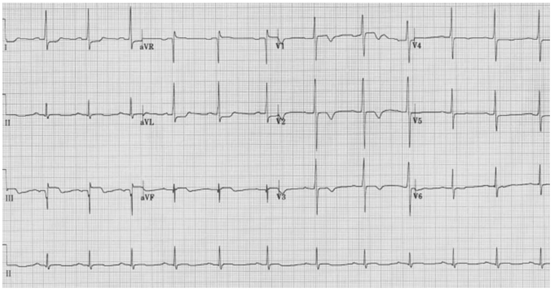
Fig. 1 Twelve-lead ECG showing sinus rhythm with ST segment elevation in leads III/aVF/aVR, ST depression in I/aVL/V5-6, and T inversion in leads V1-4. ECG: electrocardiogram.

Fig. 2 Parasternal short axis view of transthoracic echocardiography. Hypertrophic myocardium was (white arrows) localized to the anterior wall from base (A), mid-level (B), to the apex (C).
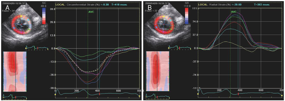
Fig. 3 Two-dimensional strain analysis. Each color represents each mid-LV segment as described in the left side of the images. The hypertrophied anterior segment, expressed as sky blue, decreased systolic deformation (strain) compared with other segments. A: circumferential strain. B: radial strain. LV: left ventricle.

Fig. 4 Consecutive static images of short axis cines {steady-state free precession (SSFP)} in diastole (A-F). Asymmetric hypertrophy of the anterior wall extended progressively from the base (A) to the apex (F) and iso-signal intensity of hypertrophied tissue compared with surrounding myocardium was seen.
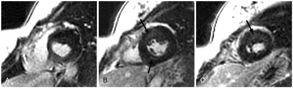
Fig. 5 Delayed contrast-enhanced MR (A-C) showed patch and linear hyperenhancement (arrows) in anterior and inferior mid-wall (different coronary artery territories).
Reference
-
1. Maron BJ. Hypertrophic cardiomyopathy: a systematic review. JAMA. 2002. 287:1308–1320.2. Wigle ED, Rakowski H, Kimball BP, Williams WG. Hypertrophic cardiomyopathy: clinical spectrum and treatment. Circulation. 1995. 92:1680–1692.3. Jeong JW. Hypertrophic cardiomyopathy. Korean Circ J. 2002. 32:7–14.4. Maron BJ, Gardin JM, Flack JM, Gidding SS, Kurosaki TT, Bild DE. Prevalence of hypertrophic cardiomyopathy in a general population of young adults: echocardiographic analysis of 4111 subjects in the CARDIA study. Circulation. 1995. 92:785–789.5. Richard P, Charron P, Carrier L, et al. Hypertrophic cardiomyopathy: distribution of disease genes, spectrum of mutations, and implications for a molecular diagnosis strategy. Circulation. 2003. 107:2227–2232.6. Klues HG, Schiffers A, Maron BJ. Phenotypic spectrum and patterns of left ventricular hypertrophy in hypertrophic cardiomyopathy. J Am Coll Cardiol. 1995. 26:1699–1708.7. Nagueh SF, Bachinski LL, Meyer D, et al. Tissue Doppler imaging consistently detects myocardial abnormalities in patients with hypertrophic cardiomyopathy and provides a novel means for an early diagnosis before and independently of hypertrophy. Circulation. 2001. 104:128–130.8. Nagueh SF, McFalls J, Meyer D, et al. Tissue Doppler imaging predicts the development of hypertrophic cardiomyopathy in subjects with subclinical disease. Circulation. 2003. 108:395–398.9. Richand V, Lafitte S, Reant P, et al. An ultrasound speckle tracking (two-dimensional strain) analysis of myocardial deformation in professional soccer players compared with healthy subjects and hypertrophic cardiomyopathy. Am J Cardiol. 2007. 100:128–132.10. Teraoka K, Hirano M, Ookubo H, et al. Delayed contrast enhancement of MRI in hypertrophic cardiomyopathy. Magn Reson Imaging. 2004. 22:155–161.11. Moon JC, McKenna WJ, McCrohon JA, Elliott PM, Smith GC, Pennell DJ. Toward clinical risk assessment in hypertrophic cardiomyopathy with gadolinium cardiovascular magnetic resonance. J Am Coll Cardiol. 2003. 41:1561–1567.12. Reichek N, Gupta D. Hypertrophic cardiomyopathy: cardiac magnetic resonance imaging changes the paradigm. J Am Coll Cardiol. 2008. 52:567–568.
- Full Text Links
-
- Actions
-
Cited
- CITED
-
- Close
- Share
-
- Similar articles
-
- A Case of Regressed Apical Hypertrophic Cardiomyopathy
- Non-invasive Assessment of Hypertrophic Cardiomyopathy
- Tips for Successful Septal Myectomy in Patients with Hypertrophic Cardiomyopathy
- A Case of Normalized Hypertrophic Cardiomyopathy after Removal of Pheochromocytoma
- Apical Hypertrophic Cardiomyopathy with Apical Aneurysm and Thrombus Diagnosed by Contrast Echocardiography
