Primary Intraosseous Meningioma in the Orbital Bony Wall: A Case Report and Review of the Literature Review
- Affiliations
-
- 1Department of Radiology, Haeundae Paik Hospital, Inje University College of Medicine, Busan, Korea. rjhrad@empal.com
- 2Department of Radiology, Busan Paik Hospital, Inje University College of Medicine, Busan, Korea.
- 3Department of Pathology, Haeundae Paik Hospital, Inje University College of Medicine, Busan, Korea.
- KMID: 1823937
- DOI: http://doi.org/10.3348/jksr.2015.72.1.68
Abstract
- Meningiomas arising outside the intracranial compartment are known as extradural meningiomas. Extradural meningiomas are rare conditions, accounting for less than 2% of all meningiomas. Primary intraosseous meningioma is used to describe a subset of extradural meningiomas arising from bone. A 46-year-old woman presented with left exophthalmos. Computed tomography and magnetic resonance images revealed an expansile bony lesion in the orbital lateral wall of the left sphenoid bone. The patient underwent craniotomy for excision of the bony lesion. Pathologic examination revealed an intraosseous meningioma.
Figure
-
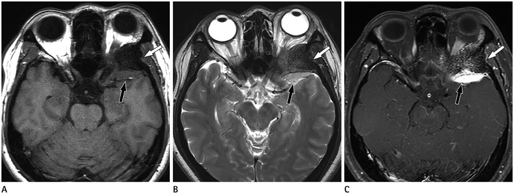
Fig. 1 A 46-year-old woman presented with left exophthalmos. Axial T1-weighted (A) and T2-weighted (B), and gadolinium-enhanced axial T1-weighted (C) MR images show an expansile bony lesion in the left lateral wall of the orbit. The intraosseous component (white arrow) is hypointense on T1- and T2-weighted images. The soft tissue component (black arrow) that represents underlying dura behind the sphenoid ridge is iso- and hyperintense on T1- and T2-weighted images with intense enhancement after administration of gadolinium.
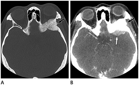
Fig. 2 CT images of a 46-year-old woman presented with left exophthalmos. A. Pre-contrast CT scan with bone widow shows an expansile and sclerotic bony lesion (arrow) in the orbital lateral wall of the left sphenoid bone. There are multiple pin-point low-attenuated foci in the bony lesion. B. Post-contrast CT images show a slightly enhancing plaque-like dural thickening (white arrow) adjacent to the bony lesion (black arrow).

Fig. 3 Histopathological feature of intraosseous hemangioma. A. Microscopic examination reveals a set of neoplastic cell lobules between bony trabecula (hematoxylin & eosin stain, × 100). B. Within a high-power field, the tumor cells have indistinct cell borders and show a whorled appearance (hematoxylin & eosin stain, × 400). C. Immunohistochemical staining for vimentin shows strong cytoplasmic reactivity (× 400).
Reference
-
1. Al-Khawaja D, Murali R, Sindler P. Primary calvarial meningioma. J Clin Neurosci. 2007; 14:1235–1239.2. Elder JB, Atkinson R, Zee CS, Chen TC. Primary intraosseous meningioma. Neurosurg Focus. 2007; 23:E13.3. Azar-Kia B, Sarwar M, Marc JA, Schechter MM. Intraosseous meningioma. Neuroradiology. 1974; 6:246–253.4. Lang FF, Macdonald OK, Fuller GN, DeMonte F. Primary extradural meningiomas: a report on nine cases and review of the literature from the era of computerized tomography scanning. J Neurosurg. 2000; 93:940–950.5. Agrawal V, Ludwig N, Agrawal A, Bulsara KR. Intraosseous intracranial meningioma. AJNR Am J Neuroradiol. 2007; 28:314–315.6. Arana E, Diaz C, Latorre FF, Menor F, Revert A, Beltrán A, et al. Primary intraosseous meningiomas. Acta Radiol. 1996; 37:937–942.7. Eisen MD, Yousem DM, Montone KT, Kotapka MJ, Bigelow DC, Bilker WB, et al. Use of preoperative MR to predict dural, perineural, and venous sinus invasion of skull base tumors. AJNR Am J Neuroradiol. 1996; 17:1937–1945.8. McGuire TP, Palme CE, Perez-Ordonez B, Gilbert RW, Sándor GK. Primary intraosseous meningioma of the calvaria: analysis of the literature and case report. Oral Surg Oral Med Oral Pathol Oral Radiol Endod. 2007; 104:e34–e41.9. Inagaki K, Otsuka F, Matsui T, Ogura T, Makino H. Effect of etidronate on intraosseous meningioma. Endocr J. 2004; 51:389–390.10. Politi M, Romeike BF, Papanagiotou P, Nabhan A, Struffert T, Feiden W, et al. Intraosseous hemangioma of the skull with dural tail sign: radiologic features with pathologic correlation. AJNR Am J Neuroradiol. 2005; 26:2049–2052.
- Full Text Links
-
- Actions
-
Cited
- CITED
-
- Close
- Share
-
- Similar articles
-
- Primary Intraosseous Calvarial Meningioma: A Case Report
- Primary Intraosseous Meningioma
- Primary Intraosseous Osteolytic Meningioma of the Skull Mimicking Scalp Mass: A Case Report and Review of Literature
- Primary Extracranial Fibrous Meningioma of the Maxillary Sinus: A Case Report and Literature Review
- Intradiploic Meningioma of Orbit Minicking Osteoma: A Case Report
