Spontaneous aortic rupture in a patient with neurofibromatosis type 1
- Affiliations
-
- 1Division of Vascular Surgery, Department of Surgery, Samsung Medical Center, Sungkyunkwan University School of Medicine, Seoul, Korea. ywkim@skku.edu
- 2Department of Interventional Radiology, Samsung Medical Center, Sungkyunkwan University School of Medicine, Seoul, Korea.
- 3Department of Pathology, Samsung Medical Center, Sungkyunkwan University School of Medicine, Seoul, Korea.
- KMID: 1820076
- DOI: http://doi.org/10.4174/jkss.2012.82.4.261
Abstract
- Neurofibromatosis type I (NF-1) is a rare autosomal dominant genetic disorder occurring in 1 in 3,000 individuals. Vasculopathy is a rarely reported finding in patients with NF-1. Here, we report a case of recurrent aortic pseudoaneurysm after endovascular aneurysm repair in a 49-year-old male patient with NF-1. On the sixth postoperative day following a successful open surgical repair of an aortic pseudoaneurysm, he developed hemoperitoneum due to a delayed rupture of the mesenteric artery branch. This was treated with endovascular coil embolization. We report the clinical features and histologic findings of this rare vascular disorder with a review of the relevant literature.
Keyword
MeSH Terms
Figure
-
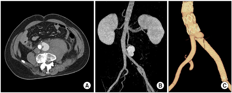
Fig. 1 (A, B) Preoperative computed tomography (CT) scan shows retroperitoneal hematoma and saccular-shaped pseudoaneurysm at left lateral wall of distal abdominal aorta at 1 cm proximal to aortic bifurcation. (C) On follow-up CT scan, small pseudoaneurysm was detected distal to aortic stent graft.

Fig. 2 (A) Microscopic section of aorta shows medial destruction with elastic fiber degeneration (upper left), thrombus in lumen, and adventitial fibrosis (Movat pentachrome stain, ×100). (B) S-100 protein-positive cells (arrow) are detected in adventitia of aorta (×200).

Fig. 3 (A) Mesenteric angiography on 6th day after open surgical repair of aortic aneurysm shows pseudoaneurysm formation at small branch of middle colic artery. (B) With coil embolization of proximal and distal segments of artery to pseudoaneurysm, bleeding from middle colic artery was successfully treated.
Reference
-
1. Friedman JM, Arbiser J, Epstein JA, Gutmann DH, Huot SJ, Lin AE, et al. Cardiovascular disease in neurofibromatosis 1: report of the NF1 Cardiovascular Task Force. Genet Med. 2002. 4:105–111.2. National Institutes of Health Consensus Development Conference Statement: neurofibromatosis. Bethesda, Md., USA, July 13-15, 1987. Neurofibromatosis. 1988. 1:172–178.3. Shimizu T, Yamazaki Y, Tomoe H, Nishino S, Toma H, Shibata T, et al. Giant retroperitoneal hematoma in a patient with von Recklinghausen's disease. Nihon Hinyokika Gakkai Zasshi. 1998. 89:846–849.4. Chew DK, Muto PM, Gordon JK, Straceski AJ, Donaldson MC. Spontaneous aortic dissection and rupture in a patient with neurofibromatosis. J Vasc Surg. 2001. 34:364–366.5. Hines GL, Lefkowitz L, Mohtashemi M. Infrarenal aortic rupture secondary to neurofibromatosis. Ann Vasc Surg. 2002. 16:784–786.6. Falcone JL, Go MR, Baril DT, Oakley GJ, Makaroun MS, Chaer RA. Vascular wall invasion in neurofibromatosis-induced aortic rupture. Vasc Endovascular Surg. 2010. 44:52–55.7. Salyer WR, Salyer DC. The vascular lesions of neurofibromatosis. Angiology. 1974. 25:510–519.8. Saitoh S, Matsuda S. Aneurysm of the major vessels in neurofibromatosis. Arch Orthop Trauma Surg. 1998. 117:110–113.9. Nonaka D, Chiriboga L, Rubin BP. Differential expression of S100 protein subtypes in malignant melanoma, and benign and malignant peripheral nerve sheath tumors. J Cutan Pathol. 2008. 35:1014–1019.10. Wolf R, Ruzicka T, Yuspa SH. Novel S100A7 (psoriasin)/S100A15 (koebnerisin) subfamily: highly homologous but distinct in regulation and function. Amino Acids. 2011. 41:789–796.
- Full Text Links
-
- Actions
-
Cited
- CITED
-
- Close
- Share
-
- Similar articles
-
- Spontaneous Hemothorax Caused by Rupture of an Intercostal Artery Aneurysm in Neurofibromatosis Type I I : A Case Report
- Lethal Hemomediastinum due to Spontaneous Rupture of an Aberrant Bronchial Artery in a Patient with Neurofibromatosis Type 1: Successful Treatment with Embolization
- Spontaneous Hemothorax in a Patient with Type I Neurofibromatosis
- Spontaneous Massive Hemothorax in a Patient with Neurofibromatosis Type 1 with Successful Transarterial Embolization
- A Case of Spontaneous Hemothorax Due to Rupture of Pseudoaneurysm in Type 1 Neurofibromatosis
