Mechanical Thrombolysis Using Coil in Acute Occlusion of Fenestrate M1 Segment
- Affiliations
-
- 1Department of Neurosurgery, Daegu Fatima Hospital, Daegu, Korea. paulyoonsoolee@hanmail.net
- 2Department of Neurosurgery, Andong General Hospital, Andong, Korea.
- KMID: 1808446
- DOI: http://doi.org/10.7461/jcen.2012.14.2.108
Abstract
- A fenestrated middle cerebral artery (MCA) is a rare congenital anomaly, and is related to interference in the normal embryonic development of the MCA. Fenestrated MCA has been regarded to have no clinical significance other than a rare event of hemorrhage from associated aneurysm. However, the fenestration within the arterial trunk can be an obstacle against thrombus migration and may be associated with a major cerebral infarction. Moreover, the presence of this anomaly can be hardly detected prior to thrombolytic procedures, and emergent treatments are proceeded without any information of anatomical configurations. Therefore, the recanalization procedures would carry a high risk of intraprocedural complications. We report a rare case of MCA territory infarction from occlusion of fenestrated M1 segment, and also introduce a safe method of mechanical thrombolysis using coil.
MeSH Terms
Figure
-
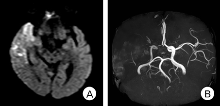
Fig. 1 (A) Diffusion-weighted image shows hyperintensity at right middle cerebral artery territory. (B) Magnetic resonance angiography demonstrates right. M1 occlusion.

Fig. 2 (A) Anterior-posterior view of right internal carotid angiogram shows right M1 occlusion. (B) Lateral view of right internal carotid angiogram reveals wide flow defect to the right middle cerebral artery territory. (C) The mechanical clot disruption was performed by simple to-and-fro maneuvers using coil. (D) Anterior-posterior view of right internal carotid angiogram shows remaining thrombus at the inferior portion of M1.

Fig. 3 (A) Follow-up angiogram after one month clearly demonstrates the anatomical contour of a fenestrated MCA. (B) The three-dimensional angiogram depicts the configuration of the fenestration with the temporopolar artery originating from distal M1.
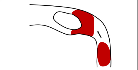
Fig. 4 The illustration shows that the fenestration within the arterial trunk may act as an obstacle against thrombus migration and can possibly be the site of occlusion.
Reference
-
1. Black SP, Ansbacher LE. Saccular aneurysm associated with segmental duplication of the basilar artery. A morphological study. J Neurosurg. 1984. 12. 61(6):1005–1008.2. Deruty R, Pelissou-Guyotat I, Mottolese C, Bognar L, Laharotte JC, Turjman F. Fenestration of the middle cerebral artery and aneurysm at the site of the fenestration. Neurol Res. 1992. 12. 14(5):421–424.
Article3. Gailloud P, Albayram S, Fasel JH, Beauchamp NJ, Murphy KJ. Angiographic and embryologic considerations in five cases of middle cerebral artery fenestration. AJNR Am J Neuroradiol. 2002. 04. 23(4):585–587.4. Jeong SK, Kwak HS, Cho YI. Middle cerebral artery fenestration in patients with cerebral ischemia. J Neurol Sci. 2008. 12. 275(1-2):181–184.
Article5. Kim MS, Hur JW, Lee JW, Lee HK. Middle cerebral artery anomalies detected by conventional angiography and magnetic resonance angiography. J Korean Neurosurg Soc. 2005. 04. 37(4):263–267.6. Nakamura H, Takada A, Hide T, Ushio Y. Fenestration of the middle cerebral artery associated with an aneurysm-case report. Neurol Med Chir (Tokyo). 1994. 08. 34(8):555–557.7. Osborn AG. Diagnostic cerebral angiography. 1999. 2nd ed. Philadelphia: Lippincott Williams and Wilkins;105135–151.8. Okudera H, Koike J, Toba Y, Kuroyanagi T, Kyoshima K, Kobayashi S. [Fenestration of the middle cerebral artery associated with cerebral infarction. Report of two cases]. Neurol Med Chir (Tokyo). 1987. 06. 27(6):559–563. Japanese.9. Paget DH. The development of the cranial arteries in the human embryo. 1948. Contrib Embryol;205–261.10. Seo BS, Lee YS, Lee HG, Lee JH, Ryu KY, Kang DG. The Clinical and Radiological Features of Patients With Aplastic or Twiglike Middle Cerebral Arteries. Neurosurgery. 2012. 06. 70(6):1472–1480.11. Sanders WP, Sorek PA, Mehta BA. Fenestration of intracranial arteries with special attention to associated aneurysms and other anomalies. AJNR Am J Neuroradiol. 1993. May-Jun. 14(3):675–680.12. Uchino A, Takase Y, Nomiyama K, Egashira R, Kudo S. Fenestration of the middle cerebral artery detected by MR angiography. Magn Reson Med Sci. 2006. 04. 5(1):51–55.
Article13. Umansky F, Dujovny M, Ausman JI, Diaz FG, Mirchandani HG. Anomalies and variations of the middle cerebral artery. A microanatomical study. Neurosurgery. 1988. 06. 22(6 Pt 1):1023–1027.
- Full Text Links
-
- Actions
-
Cited
- CITED
-
- Close
- Share
-
- Similar articles
-
- Effect of Intravenous Thrombolysis Prior to Mechanical Thrombectomy According to the Location of M1 Occlusion
- Mechanical thrombectomy for acute ischemic stroke with occlusion of the M2 segment of the middle cerebral artery: A literature review
- Intra-arterial Thrombolysis for Central Retinal Artery Occlusion after the Coil Embolization of Paraclinoid Aneurysm
- Interventional Recanalization Treatment of Acute Ischemic Stroke
- Endovascular Therapy for Acute Basilar Artery Occlusion: Comparison between Patients with and without Underlying Intracranial Atherosclerotic Stenosis
