J Cardiovasc Ultrasound.
2015 Jun;23(2):118-118. 10.4250/jcu.2015.23.2.118.
Giant Carotid Pseudoaneurysm
- Affiliations
-
- 1Division of Neurology, Department of Medicine, National University Health System, Singapore. leonard_ll_yeo@nuhs.edu.sg
- 2Department of Cardiac, Thoracic and Vascular Surgery, National University Health System, Singapore.
- 3Department of Diagnostic Imaging, National University Health System, Singapore.
- 4Yong Loo Lin School of Medicine, National University of Singapore, Singapore.
- KMID: 1806924
- DOI: http://doi.org/10.4250/jcu.2015.23.2.118
Abstract
- No abstract available.
Keyword
MeSH Terms
Figure
-
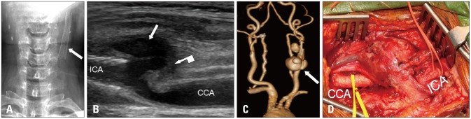
Fig. 1 Neck X-ray showing a long left carotid stent (arrow) (A). Cervical duplex showing the pseudoaneurysm originating from distal common carotid artery (straight arrow, B). Computed tomography angiography of the neck (C). The lobulated pseudoaneurysm (arrow, C) wrapped around the carotid system (D) multiple thrombi on the wall of pseudoaneurysm (bent arrow, B) confirmed during surgery. CCA: common carotid artery, ICA: internal carotid artery.
Reference
-
1. Saad NE, Saad WE, Davies MG, Waldman DL, Fultz PJ, Rubens DJ. Pseudoaneurysms and the role of minimally invasive techniques in their management. Radiographics. 2005; 25(Suppl 1):S173–S189. PMID: 16227490.
- Full Text Links
-
- Actions
-
Cited
- CITED
-
- Close
- Share
-
- Similar articles
-
- Conjoined Stent Technique for Radiation Induced Long Segment Carotid Stenosis and Pseudoaneurysm
- Symptomatic Post Endarterectomy Common Carotid Artery Pseudoaneurysm Treated with Combination of Flow Diverter Implantation and Carotid Stenting
- Traumatic Pseudoaneurysm of the External and Internal Carotid Artery Presenting as Epistaxis: Case Report
- Parent artery occlusion of a giant internal carotid artery pseudoaneurysm-related direct carotid cavernous fistula: A case report
- Endovascualr Treatment for Traumatic Giant Pseudoaneurysm of Internal Carotid Artery
