Measurements of simulated periodontal bone defects in inverted digital image and film-based radiograph: an in vitro study
- Affiliations
-
- 1Department of Diagnosis and Surgery, Araraquara Dental School, Universidade Estadual Paulista, Sao Paulo, Brazil. molon.foar@yahoo.com.br
- 2Barretos Dental School, Barretos Educational Fundation, Sao Paulo, Brazil.
- 3Department of Social Dentistry, Araraquara Dental School, Universidade Estadual Paulista, Sao Paulo, Brazil.
- KMID: 1806791
- DOI: http://doi.org/10.5624/isd.2012.42.4.243
Abstract
- PURPOSE
This study was performed to compare the inverted digital images and film-based images of dry pig mandibles to measure the periodontal bone defect depth.
MATERIALS AND METHODS
Forty 2-wall bone defects were made in the proximal region of the premolar in the dry pig mandibles. The digital and conventional radiographs were taken using a Schick sensor and Kodak F-speed intraoral film. Image manipulation (inversion) was performed using Adobe Photoshop 7.0 software. Four trained examiners made all of the radiographic measurements in millimeters a total of three times from the cementoenamel junction to the most apical extension of the bone loss with both types of images: inverted digital and film. The measurements were also made in dry mandibles using a periodontal probe and digital caliper. The Student's t-test was used to compare the depth measurements obtained from the two types of images and direct visual measurement in the dry mandibles. A significance level of 0.05 for a 95% confidence interval was used for each comparison.
RESULTS
There was a significant difference between depth measurements in the inverted digital images and direct visual measurements (p>|t|=0.0039), with means of 6.29 mm (IC95%:6.04-6.54) and 6.79 mm (IC95%:6.45-7.11), respectively. There was a non-significant difference between the film-based radiographs and direct visual measurements (p>|t|=0.4950), with means of 6.64mm(IC95%:6.40-6.89) and 6.79mm(IC95%:6.45-7.11), respectively.
CONCLUSION
The periodontal bone defect measurements in the inverted digital images were inferior to film-based radiographs, underestimating the amount of bone loss.
MeSH Terms
Figure
-

Fig. 1 A. Film-based image shows the distance from the cementoenamel junction to the deepest region of the defect. B. Inverted digital image shows the distance from the cementoenamel junction to the deepest region of the defect.
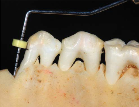
Fig. 2 Direct bone-defect measurement in a dry pig mandible.

Fig. 3 Mean (m) and 95% confidence limits (LS: upper limit; LI: lower limit) of the bone defect depth (in mm) according to the method (M: direct visual measurements; D: inverted digital image).
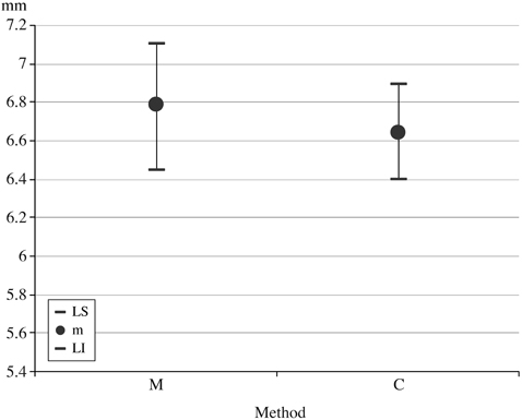
Fig. 4 Mean (m) and 95% confidence limits (LS: upper limit; LI: lower limit) of the bone defect depth (in mm) according to the method (M: direct visual measurements; C: film-based image).
Cited by 1 articles
-
Effect of digital noise reduction on the accuracy of endodontic file length determination
Mojdeh Mehdizadeh, Abbas Ali Khademi, Ali Shokraneh, Nastaran Farhadi
Imaging Sci Dent. 2013;43(3):185-190. doi: 10.5624/isd.2013.43.3.185.
Reference
-
1. Scaf G, Morihisa O, Loffredo Lde C. Comparison between inverted and unprocessed digitized radiographic imaging in periodontal bone loss measurements. J Appl Oral Sci. 2007. 15:492–494.
Article2. Scaf G, Sakakura CE, Kalil PF, Dearo de Morais JA, Loffredo LC, Wenzel A. Comparison of simulated periodontal bone defect depth measured in digital radiographs in dedicated and non-dedicated software systems. Dentomaxillofac Radiol. 2006. 35:422–425.
Article3. Tihanyi D, Gera I, Eickholz P. Influence of individual brightness and contrast adjustment on accuracy of radiographic measurements of infrabony defects. Dentomaxillofac Radiol. 2011. 40:177–183.
Article4. de Molon RS, Sakakura CE, Morais-Camillo JA, de Almeida Junior PC, de Castro Monteiro Loffredo L, Scaf G. Comparison between embossed digital imaging and unprocessed film-based radiography in detecting periodontal bone defects: an in vitro study. Oral Radiol. 2012. 28:95–100.
Article5. Borg E, Gröndahl K, Gröndahl HG. Marginal bone level buccal to mandibular molars in digital radiographs from charge-coupled device and storage phosphor systems. An in vitro study. J Clin Periodontol. 1997. 24:306–312.6. Eickholz P, Hausmann E. Accuracy of radiographic assessment of interproximal bone loss in intrabony defects using linear measurements. Eur J Oral Sci. 2000. 108:70–73.
Article7. de Morais JA, Sakakura CE, Loffredo LC, Scaf G. Accuracy of zoomed digital image in the detection of periodontal bone defect: in vitro study. Dentomaxillofac Radiol. 2006. 35:139–142.8. Sakakura CE, Loffredo Lde C, Scaf G. Diagnostic agreement of conventional and inverted scanned panoramic radiographs in the detection of the mandibular canal and the mental foramen. J Oral Implantol. 2004. 30:2–6.
Article9. Tyndall DA, Ludlow JB, Platin E, Nair M. A comparison of Kodak Ektaspeed Plus film and the Siemens Sidexis digital imaging system for caries detection using receiver operating characteristic analysis. Oral Surg Oral Med Oral Pathol Oral Radiol Endod. 1998. 85:113–118.
Article10. Isidor S, Faaborg-Andersen M, Hintze H, Kirkevang LL, Frydenberg M, Haiter-Neto F, et al. Effect of monitor display on detection of approximal caries lesions in digital radiographs. Dentomaxillofac Radiol. 2009. 38:537–541.
Article11. Hellén-Halme K, Petersson A, Warfvinge G, Nilsson M. Effect of ambient light and monitor brightness and contrast settings on the detection of approximal caries in digital radiographs: an in vitro study. Dentomaxillofac Radiol. 2008. 37:380–384.12. Eickholz P, Riess T, Lenhard M, Hassfeld S, Staehle HJ. Digital radiography of interproximal bone loss; validity of different filters. J Clin Periodontol. 1999. 26:294–300.
Article13. Nair MK, Ludlow JB, Tyndall DA, Platin E, Denton G. Periodontitis detection efficacy of film and digital images. Oral Surg Oral Med Oral Pathol Oral Radiol Endod. 1998. 85:608–612.
Article14. Wolf B, Von Bethlenfalvy E, Hassfeld S, Staehle HJ, Eickholz P. Reliability of assessing interproximal bone loss by digital radiography: intrabony defects. J Clin Periodontol. 2001. 28:869–878.
Article15. Eickholz P, Kim TS, Benn DK, Staehle HJ. Validity of radiographic measurement of interproximal bone loss. Oral Surg Oral Med Oral Pathol Oral Radiol Endod. 1998. 85:99–106.
Article16. Furkart AJ, Dove SB, McDavid WD, Nummikoski P, Matteson S. Direct digital radiography for the detection of periodontal bone lesions. Oral Surg Oral Med Oral Pathol. 1992. 74:652–660.
Article
- Full Text Links
-
- Actions
-
Cited
- CITED
-
- Close
- Share
-
- Similar articles
-
- An experimental study on the readability of digital images in the furcal bone defects
- Digital imaging of film-based cephalograms using a digital camera
- Detectability of Ektaspeed Puls Film, Digitized and Digora Images for artificial periapical bone lesions
- Experimental study of alveolar bone wall defects using direct digital radiography
- The Comparative Study of Alveolar Bone Level and Root Form of the Mandibular Molar on Radiographic Image and Clinical Examination
