Yonsei Med J.
2010 Jul;51(4):590-593. 10.3349/ymj.2010.51.4.590.
Diffusion Tensor Imaging of Heterotopia: Changes of Fractional Anisotropy during Radial Migration of Neurons
- Affiliations
-
- 1Department of Radiology and Research Institute of Radiological Science, Yonsei University College of Medicine, Seoul, Korea. slee@yuhs.ac
- KMID: 1805200
- DOI: http://doi.org/10.3349/ymj.2010.51.4.590
Abstract
- PURPOSE
Diffusion tensor imaging provides better understanding of pathophysiology of congenital anomalies, involving central nervous system. This study was aimed to specify the pathogenetic mechanism of heterotopia, proved by diffusion tensor imaging, and establish new findings of heterotopia on fractional anisotropy maps.
MATERIALS AND METHODS
Diffusion-weighted imaging data from 11 patients (M : F = 7 : 4, aged from 1 to 22 years, mean = 12.3 years) who visited the epilepsy clinic and received a routine seizure protocol MRI exam were retrospectively analyzed. Fractional anisotropy (FA) maps were generated from diffusion tensor imaging of 11 patients with heterotopia. Regions of interests (ROI) were placed in cerebral cortex, heterotopic gray matter and deep gray matter, including putamen. ANOVA analysis was performed for comparison of different gray matter tissues.
RESULTS
Heterotopic gray matter showed signal intensities similar to normal gray matter on T1 and T2 weighted MRI. The measured FA of heterotopic gray matter was higher than that of cortical gray matter (0.236 +/- 0.011 vs. 0.169 +/- 0.015, p < 0.01, one way ANOVA), and slightly lower than that of deep gray matter (0.236 +/- 0.011 vs. 0.259 +/- 0.016, p < 0.01).
CONCLUSION
Increased FA of heterotopic gray matter suggests arrested neuron during radial migration and provides better understanding of neurodevelopment.
Figure
-
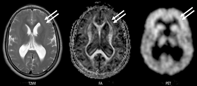
Fig. 1 Twenty two year old male with intractable epilepsy. Note linear band of gray matter tissue with similar signal intensity as cortex and high FA values (double arrows). On PET scan, this heterotopic gray matter also shows high glucose metabolism than white matter (double arrows). FA, fractional anisotropy.

Fig. 2 Fractional anisotropy (FA) of gray matter tissues. Deep gray matter (DGM) is higher than heterotopic gray matter (HGM) and cortical gray matter (CGM). Note FA of HGM is also significantly higher than CGM.
Reference
-
1. Livingston JH, Aicardi J. Unusual MRI appearance of diffuse subcortical heterotopia or "double cortex" in two children. J Neurol Neurosurg Psychiatry. 1990. 53:617–620.
Article2. Gallucci M, Bozzao A, Curatolo P, Splendiani A, Cifani A, Passariello R. MR imaging of incomplete band heterotopia. AJNR Am J Neuroradiol. 1991. 12:701–702.3. Bourgeois JA, Nisenbaum J, Drexler KG, Dobbins KM, Hall MJ. A case of subcortical grey matter heterotopia presenting as bipolar disorder. Compr Psychiatry. 1992. 33:407–410.
Article4. Kamuro K, Tenokuchi Y. Familial periventricular nodular heterotopia. Brain Dev. 1993. 15:237–241.
Article5. Bauer J, Elger CE. Band heterotopia: a rare cause of generalized epileptic seizures. Seizure. 1994. 3:153–155.6. Eriksson SH, Symms MR, Rugg-Gunn FJ, Boulby PA, Wheeler-Kingshott CA, Barker GJ, et al. Exploring white matter tracts in band heterotopia using diffusion tractography. Ann Neurol. 2002. 52:327–334.
Article7. Huttenlocher PR, Taravath S, Mojtahedi S. Periventricular heterotopia and epilepsy. Neurology. 1994. 44:51–55.
Article8. Gleeson JG, Walsh CA. Neuronal migration disorders: from genetic diseases to developmental mechanisms. Trends Neurosci. 2000. 23:352–359.
Article9. Barkovich AJ. Morphologic characteristics of subcortical heterotopia: MR imaging study. AJNR Am J Neuroradiol. 2000. 21:290–295.10. Pinard J, Feydy A, Carlier R, Perez N, Pierot L, Burnod Y. Functional MRI in double cortex: functionality of heterotopia. Neurology. 2000. 54:1531–1533.
Article11. Spreer J, Martin P, Greenlee MW, Wohlfarth R, Hammen A, Arnold SM, et al. Functional MRI in patients with band heterotopia. Neuroimage. 2001. 14:357–365.
Article12. Villani F, Vitali P, Scaioli V, Rodriguez G, Rosa M, Granata T, et al. Subcortical nodular heterotopia: a functional MRI and somatosensory evoked potentials study. Neurol Sci. 2004. 25:225–229.
Article13. Castillo M, Kwock L, Scatliff J, Gudeman S, Greenwood R. Proton MR spectroscopic characteristics of a presumed giant subcortical heterotopia. AJNR Am J Neuroradiol. 1993. 14:426–429.14. Mai R, Tassi L, Cossu M, Francione S, Lo Russo G, Garbelli R, et al. A neuropathological, stereo-EEg, and MRI study of subcortical band heterotopia. Neurology. 2003. 60:1834–1838.
Article15. Nucifora PG, Verma R, Lee SK, Melhem ER. Diffusion-tensor MR imaging and tractography: exploring brain microstructure and connectivity. Radiology. 2007. 245:367–384.
Article
- Full Text Links
-
- Actions
-
Cited
- CITED
-
- Close
- Share
-
- Similar articles
-
- Image Reconstruction of Eigenvalue of Diffusion Principal Axis Using Diffusion Tensor Imaging
- The Changes in Axial and Radial Diffusivity in a Patient with Clinically Mild Encephalitis/Encephalopathy with a Reversible Splenial Lesion
- Diffusion Metrics as a Potential Prognostic Biomarker in Cervical Myelopathy
- Diffusion Tensor Imaging Findings in Two Cases of Internal Capsular Genu Infarction
- Analysis of Diffuse Axonal Injury Using Diffusion Tensor Imaging in Traumatic Brain Injury Patients
