Yonsei Med J.
2014 Sep;55(5):1447-1449. 10.3349/ymj.2014.55.5.1447.
Silicon Dioxide Particles Deposited in Vessels and Cartilage of the Femoral Head
- Affiliations
-
- 1Department of Intensive Care Unit, Second Xiangya Hospital, Central South University, Hunan, P. R. China.
- 2Department of Urinary Surgery, Second Xiangya Hospital, Central South University, Hunan, P. R. China.
- 3Department of Orthopedics, Second Xiangya Hospital, Central South University, Hunan, P. R. China. xyeypd@163.com
- KMID: 1799516
- DOI: http://doi.org/10.3349/ymj.2014.55.5.1447
Abstract
- Silicosis had been considered for decades as an illness with manifestations of lung fibrosis due to inhalation of overconcentrated SiO2 dust. To the best of our knowledge, studies have yet to report SiO2 deposits in any other tissues and organs. In the present case, while performing bilateral artificial total hip arthroplasty for one patient, we found that the articular cartilage of the bilateral femoral head was black. Therefore, specimens thereof were sent for pathological examination. Pathological examination (immunohistochemistry) and polarized light microscopy revealed the presence of considerable brown, acicular, rhombic, and crumb-like crystals. The crystals were mainly composed of SiO2. SiO2 could deposit in vessels and femoral head cartilage via blood circulation.
Keyword
MeSH Terms
Figure
-
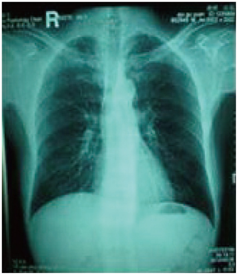
Fig. 1 Radiograph of the lungs: lung markings of both lungs are increased and thick; mottled, unclear edges and diffuse distribution of high density material in the bilateral lungs are visible. The radiograph is consistent with early silicosis.
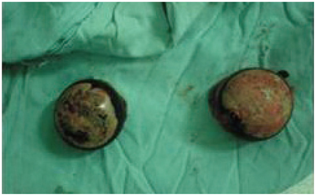
Fig. 2 Excised bilateral femoral head.
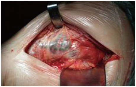
Fig. 3 Black substances in vessels of the left greater trochanter.
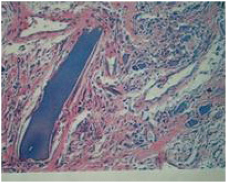
Fig. 4 Pathological examination (immunohistochemistry) and polarized light microscopy: chronic synovitis and proliferation of granuloma are visible under a microscope. Note the considerable amounts of brownish acicular, rhombic, and crumb-like crystals. At their edges, giant cell reactions of foreign-bodies are seen. Crystals mainly comprised SiO2 (H&E ×100).
Reference
-
1. Ambrosi L. [Histologic aspects of the spleen in silicosis patients]. Med Lav. 1966; 57:10–19.2. Ambrosi L. [Histologic aspects of the liver in silicosis patients]. Med Lav. 1965; 56:795–804.3. Ambrosi L. [Histologic aspects of the kidney in silicosis]. Med Lav. 1965; 56:716–726.4. Ambrosi L. [Histological aspects of the heart in silicotic patients]. Med Lav. 1966; 57:104–110.5. Carmichael GP Jr, Targoff C, Pintar K, Lewin KJ. Hepatic silicosis. Am J Clin Pathol. 1980; 73:720–722.
Article6. Autio L, Turunen M, Lahtiharju A. Experimental silica cirrhosis in dog. Ann Med Exp Biol Fenn. 1964; 42:173–176.7. Roperto F, Troncone A, Tranquillo A, Galati P. Extrapulmonary silicosis in two water buffaloes. J Comp Pathol. 1995; 112:97–103.
Article8. Montaldo G, Onnis C, Montaldo S. [Pneumosilicosis]. Arch De Vecchi Anat Patol. 1976; 61:487–495.
- Full Text Links
-
- Actions
-
Cited
- CITED
-
- Close
- Share
-
- Similar articles
-
- Out-patient visits for respiratory diseases and yellow sand phenomena
- Histological and Histochemical Study of the Acetabular Articular Cartilage in Avascular Necrosis of Femoral Head
- Silica Complexed with Fe3+ Does not Influence Pulmonary Inflammation and Injury
- Histopathological changes in rat lung instilled with natural coal and free silica dust
- Cement dust and environmental diseases
