Changes in Spinal Canal Diameter and Vertebral Body Height with Age
- Affiliations
-
- 1Department of Neurosurgery, Gangnam Severance Hospital, Spine and Spinal Cord Institute, Yonsei University College of Medicine, Seoul, Korea. spinepjy@yuhs.ac
- KMID: 1798149
- DOI: http://doi.org/10.3349/ymj.2013.54.6.1498
Abstract
- PURPOSE
All structures of the spine, including the spinal canal, change continuously with age. The purpose of this study was to determine how the spinal canal of the lumbar spine changes with age. The L4/5 is the most common site of spinal stenosis and has the largest flexion-extension motion, whereas the T5/6 has the least motion. Therefore, we measured the spinal canal diameter and vertebral body height at T5, T6, L4, and L5 with age.
MATERIALS AND METHODS
This was a retrospective study of aged 40 to 77 years. We reviewed whole spine sagittal MRIs of 370 patients with lumbar spinal stenosis (LSS) (Group 2) and 166 herniated cervical disc (HCD) (Group 1). Each group was divided into four age groups, and demographic parameters (age, gender, height, weight, BMI), the mid-spinal canal diameter, and mid-vertebrae height at T5, T6, L4, L5 were compared. Within- and between-group comparisons were made to evaluate changes by age and correlations were carried out to evaluate the relationships between all parameters.
RESULTS
Height, weight, and all radiologic parameters were significantly lower in Group 2 than Group 1. Group 1 did not show any differences, when based on age, but in Group 2, height, weight, and T6, L4, and L5 height were significantly decreased in patients in their 70's than patients in their 40's, except for spinal canal diameter. Age was associated with all parameters except spinal canal diameter.
CONCLUSION
Vertebral height decreased with age, but spinal canal diameter did not change in patients with either LSS or HCD. Mid-spinal canal diameter was not affected by aging.
MeSH Terms
Figure
-
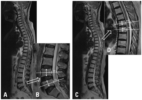
Fig. 1 Radiologic parameters of whole spine sagittal MRI. From the whole spine sagittal MRI, we selected the largest spinal canal image at the lumbar spine (A). The anteroposterior diameter of the spinal canal at the mid-portion of the vertebral body (white line) and mid-vertebra body height (white arrow) at L4 and L5 was measured (B). From the whole spine sagittal MRI, we selected the largest spinal canal image at the thoracic spine (C). The anteroposterior diameter of the spinal canal at the mid-portion of the vertebral body (white line) and mid-vertebra body height (white arrow) at T5 and T6 was measured (D).
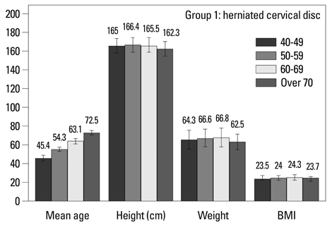
Fig. 2 Demographic characteristics in Group 1 (herniated cervical disc).
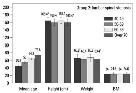
Fig. 3 Demographic characteristics in Group 2 (lumbar spinal stenosis). Comparisons between 40-49 year olds and over 70 year olds by Student's t-test, *p<0.01 and †p<0.05.
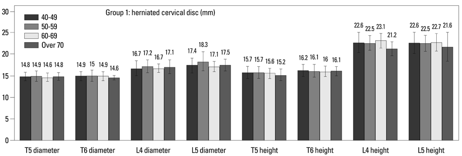
Fig. 4 Spinal canal diameters and vertebral body heights in Group 1 (herniated cervical disc).

Fig. 5 Spinal canal diameters and vertebral body heights in Group 2 (lumbar spinal stenosis). Comparisons between 40-49 year olds and over 70 year olds by Student's t-test, *p<0.01.
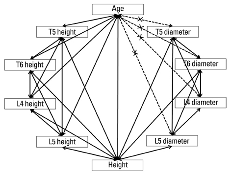
Fig. 6 Correlations between all parameters. Age was significantly correlated with all demographic characteristics and vertebral body heights, but did not correlate with spinal canal diameter.
Reference
-
1. Shao Z, Rompe G, Schiltenwolf M. Radiographic changes in the lumbar intervertebral discs and lumbar vertebrae with age. Spine (Phila Pa 1976). 2002; 27:263–268.
Article2. Ericksen MF. Some aspects of aging in the lumbar spine. Am J Phys Anthropol. 1976; 45(3 pt. 2):575–580.
Article3. Vernon-Roberts B, Moore RJ, Fraser RD. The natural history of age-related disc degeneration: the influence of age and pathology on cell populations in the L4-L5 disc. Spine (Phila Pa 1976). 2008; 33:2767–2773.4. Twomey L, Taylor J. Age changes in the lumbar spinal and intervertebral canals. Paraplegia. 1988; 26:238–249.
Article5. Abbas J, Hamoud K, May H, Hay O, Medlej B, Masharawi Y, et al. Degenerative lumbar spinal stenosis and lumbar spine configuration. Eur Spine J. 2010; 19:1865–1873.
Article6. Arnoldi CC, Brodsky AE, Cauchoix J, Crock HV, Dommisse GF, Edgar MA, et al. Lumbar spinal stenosis and nerve root entrapment syndromes. Definition and classification. Clin Orthop Relat Res. 1976; 4–5.7. Bartolozzi P, Salvi M, Misasi M. The diagnosis of lumbar stenosis. Chir Organi Mov. 1992; 77:15–18.8. Eisenstein S. The morphometry and pathological anatomy of the lumbar spine in South African negroes and caucasoids with specific reference to spinal stenosis. J Bone Joint Surg Br. 1977; 59:173–180.
Article9. Katz JN, Dalgas M, Stucki G, Lipson SJ. Diagnosis of lumbar spinal stenosis. Rheum Dis Clin North Am. 1994; 20:471–483.
Article10. Singh K, Samartzis D, Vaccaro AR, Nassr A, Andersson GB, Yoon ST, et al. Congenital lumbar spinal stenosis: a prospective, control-matched, cohort radiographic analysis. Spine J. 2005; 5:615–622.
Article11. Kirkaldy-Willis WH, Wedge JH, Yong-Hing K, Reilly J. Pathology and pathogenesis of lumbar spondylosis and stenosis. Spine (Phila Pa 1976). 1978; 3:319–328.
Article12. Kirkaldy-Willis WH, Paine KW, Cauchoix J, McIvor G. Lumbar spinal stenosis. Clin Orthop Relat Res. 1974; 30–50.13. Kirkaldy-Willis WH, Wedge JH, Yong-Hing K, Tchang S, de Korompay V, Shannon R. Lumbar spinal nerve lateral entrapment. Clin Orthop Relat Res. 1982; 171–178.
Article14. Panjabi MM, White AA 3rd. Basic biomechanics of the spine. Neurosurgery. 1980; 7:76–93.
Article15. Panjabi MM, Goel V, Oxland T, Takata K, Duranceau J, Krag M, et al. Human lumbar vertebrae. Quantitative three-dimensional anatomy. Spine (Phila Pa 1976). 1992; 17:299–306.16. Panjabi MM, Takata K, Goel V, Federico D, Oxland T, Duranceau J, et al. Thoracic human vertebrae. Quantitative three-dimensional anatomy. Spine (Phila Pa 1976). 1991; 16:888–901.
Article17. White AA 3rd, Panjabi MM. The basic kinematics of the human spine. A review of past and current knowledge. Spine (Phila Pa 1976). 1978; 3:12–20.18. Amundsen T, Weber H, Nordal HJ, Magnaes B, Abdelnoor M, Lilleâs F. Lumbar spinal stenosis: conservative or surgical management?: a prospective 10-year study. Spine (Phila Pa 1976). 2000; 25:1424–1435.19. Ericksen MF. Aging in the lumber spine. III. L5. Am J Phys Anthropol. 1978; 48:247–250.20. Ericksen MF. Aging in the lumbar spine. II. L1 and L2. Am J Phys Anthropol. 1978; 48:241–245.
Article21. Masharawi Y, Salame K, Mirovsky Y, Peleg S, Dar G, Steinberg N, et al. Vertebral body shape variation in the thoracic and lumbar spine: characterization of its asymmetry and wedging. Clin Anat. 2008; 21:46–54.
Article22. Postacchini F. Management of lumbar spinal stenosis. J Bone Joint Surg Br. 1996; 78:154–164.
Article23. Masharawi Y, Salame K. Shape variation of the neural arch in the thoracic and lumbar spine: characterization and relationship with the vertebral body shape. Clin Anat. 2011; 24:858–867.
Article
- Full Text Links
-
- Actions
-
Cited
- CITED
-
- Close
- Share
-
- Similar articles
-
- Mid-sagittal canal diameter and vertebral body/canal ratio of the cervical spine in Koreans
- Analysis of Factors Related to Neurological Deficit in Thoracolumbar Fractures
- Computed tomographic evaluation of cervical vertebral canal and spinal cord morphometry in normal dogs
- Preliminary Report of Percutaneous Vertebroplasty for the Treatment of the Burst Fractures with Spinal Canal Encroachment
- Pavlov’s Ratio of the Cervical Spine in a Korean Population: A Comparative Study by Age in Patients with Minor Trauma without Neurologic Symptoms
