Visibility of Sutures of the Orbit and Periorbital Region Using Multidetector Computed Tomography
- Affiliations
-
- 1Department of Diagnostic Radiology, Martin-Luther-University Halle-Wittenberg, 06120 Halle (Saale), Germany. hgufler@gmx.de
- KMID: 1794653
- DOI: http://doi.org/10.3348/kjr.2014.15.6.802
Abstract
OBJECTIVE
Knowledge of cranial suture morphology is crucial in emergency medicine, forensic medicine, and maxillofacial reconstructive surgery. This study assessed the visibility of sutures of the orbit and periorbital region on multidetector computed tomography.
MATERIALS AND METHODS
Multidetector computed tomography scans of 200 patients (127 males, 73 females; mean age 51.3 years; range, 6-92 years) were evaluated retrospectively. The slice thicknesses varied from 0.5 to 1 mm, and the tube current from 25 to 370 mAs, depending on the CT indication. The visibility of sutures was estimated according to a 4-point scale from "not visible" to "well visible". The chi-squared test was used to test the association of the visibility of sutures with the slice thickness, tube current, and age of patients. Statistical significance was assumed at p < 0.05.
RESULTS
Overall, best visibility was found for the sutura frontozygomatica (98%), sutura frontonasalis (88.5%), and sutura sphenozygomatica (71.5%), followed by the sutura zygomaticomaxillaris (65.8%), sutura temporozygomatica (41.8%), sutura frontomaxillaris (44.5%), and sutura sphenofrontalis (31%). Poor visibility was found for the sutura frontolacrimalis (16.8%) and sutura frontoethmoidalis (1.3%). The sutura ethmoidomaxillaris, sutura lacrimomaxillaris, and sutura ethmoidolacrimalis were not visible.
CONCLUSION
Although the sutures of the superior, lateral, and inferior orbit are well visible, those of the medial orbit are poorly visible on CT scans.
MeSH Terms
Figure
-
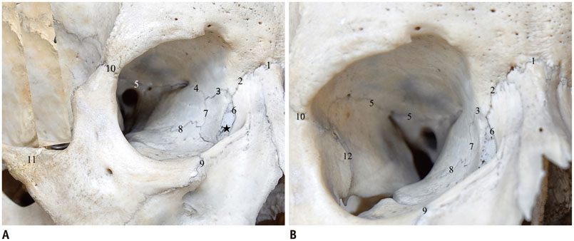
Fig. 1 Two different views on sutures of right orbit and periorbital region. A. Sutures of medial orbital wall. B. Sutures of lateral orbital wall. 1 = sutura frontonasalis, 2 = sutura frontomaxillaris, 3 = sutura frontolacrimalis, 4 = sutura frontoethmoidalis, 5 = sutura sphenofrontalis, 6 = sutura lacrimomaxillaris, 7 = sutura ethmoidolacrimalis, 8 = sutura ethmoidomaxillaris, 9 = sutura zygomaticomaxillaris, 10 = sutura frontozygomatica, 11 = sutura temporozygomatica, 12 = sutura sphenozygomatica. Star = fossa lacrimalis

Fig. 2 Sutura frontozygomatica (arrow), coronal multiplanar reconstruction.
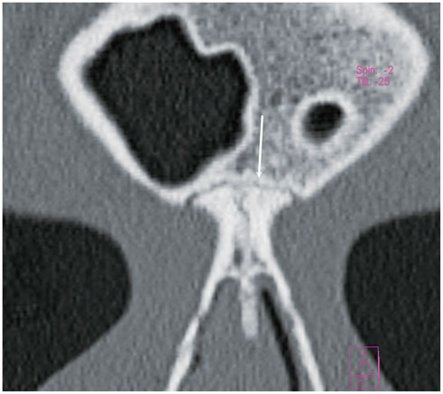
Fig. 3 Sutura frontonasalis (arrow), coronal multiplanar reconstruction.

Fig. 4 Sutura sphenozygomatica (arrow), oblique multiplanar reconstruction.
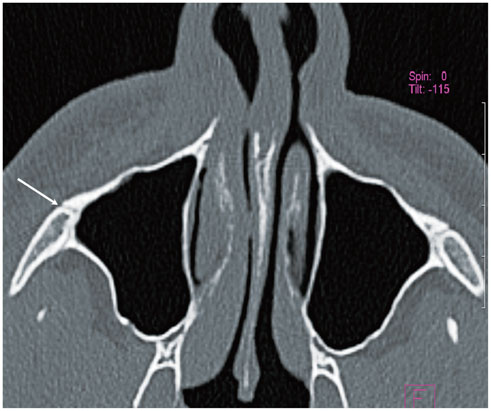
Fig. 5 Sutura zygomaticomaxillaris (arrow), axial CT.

Fig. 6 Sutura temporozygomatica (arrow), axial multiplanar reconstruction.
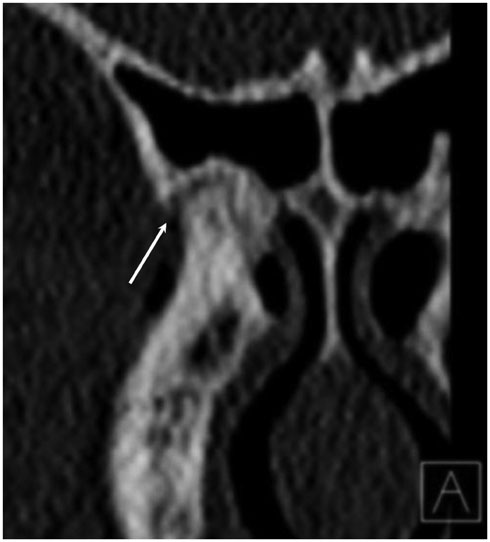
Fig. 7 Sutura frontomaxillaris (arrow), angulated coronal multiplanar reconstruction.

Fig. 8 Sutura sphenofrontalis (solid arrow) and depiction of sutura sphenozygomatica (dashed arrow), angulated coronal multiplanar reconstruction.

Fig. 9 Sutura frontolacrimalis (arrow), coronal multiplanar reconstruction.
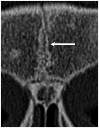
Fig. 10 Sutura frontalis persistens (arrow), axial scan.
Reference
-
1. Harth S, Obert M, Ramsthaler F, Reuss C, Traupe H, Verhoff MA. Ossification degrees of cranial sutures determined with flat-panel computed tomography: narrowing the age estimate with extrema. J Forensic Sci. 2010; 55:690–694.2. Madeline LA, Elster AD. Suture closure in the human chondrocranium: CT assessment. Radiology. 1995; 196:747–756.3. Vannier MW, Pilgram TK, Marsh JL, Kraemer BB, Rayne SC, Gado MH, et al. Craniosynostosis: diagnostic imaging with three-dimensional CT presentation. AJNR Am J Neuroradiol. 1994; 15:1861–1869.4. Sim SY, Yoon SH, Kim SY. Quantitative analysis of developmental process of cranial suture in korean infants. J Korean Neurosurg Soc. 2012; 51:31–36.5. Choudhary AK, Jha B, Boal DK, Dias M. Occipital sutures and its variations: the value of 3D-CT and how to differentiate it from fractures using 3D-CT? Surg Radiol Anat. 2010; 32:807–816.6. Bademci G, Kendi T, Agalar F. Persistent metopic suture can mimic the skull fractures in the emergency setting? Neurocirugia (Astur). 2007; 18:238–224.7. Furuya Y, Edwards MS, Alpers CE, Tress BM, Ousterhout DK, Norman D. Computerized tomography of cranial sutures. Part 1: comparison of suture anatomy in children and adults. J Neurosurg. 1984; 61:53–55.8. Tack D, Widelec J, De Maertelaer V, Bailly JM, Delcour C, Gevenois PA. Comparison between low-dose and standard-dose multidetector CT in patients with suspected chronic sinusitis. AJR Am J Roentgenol. 2003; 181:939–944.9. Wang Q, Strait DS, Dechow PC. Fusion patterns of craniofacial sutures in rhesus monkey skulls of known age and sex from Cayo Santiago. Am J Phys Anthropol. 2006; 131:469–485.10. Dorandeu A, Coulibaly B, Piercecchi-Marti MD, Bartoli C, Gaudart J, Baccino E, et al. Age-at-death estimation based on the study of frontosphenoidal sutures. Forensic Sci Int. 2008; 177:47–51.11. Todd TW, Lyon DW Jr. Cranial suture closure. Its progress and age relationship. Part II. Ectocranial closure in adult males of white stock. Am J Phys Anthropol. 1925; 8:23–45.12. Mann RW, Jantz RL, Bass WM, Willey PS. Maxillary suture obliteration: a visual method for estimating skeletal age. J Forensic Sci. 1991; 36:781–791.13. Beauthier JP, Lefevre P, Meunier M, Orban R, Polet C, Werquin JP, et al. Palatine sutures as age indicator: a controlled study in the elderly. J Forensic Sci. 2010; 55:153–158.14. Ajmani ML, Mittal RK, Jain SP. Incidence of the metopic suture in adult Nigerian skulls. J Anat. 1983; 137(Pt 1):177–183.15. Agarwal SK, Malhotra VK, Tewari SP. Incidence of the metopic suture in adult Indian crania. Acta Anat (Basel). 1979; 105:469–447.16. Baaten PJ, Haddad M, Abi-Nader K, Abi-Ghosn A, Al-Kutoubi A, Jurjus AR. Incidence of metopism in the Lebanese population. Clin Anat. 2003; 16:148–151.17. Eroğlu S. The frequency of metopism in Anatolian populations dated from the Neolithic to the first quarter of the 20th century. Clin Anat. 2008; 21:471–478.18. Lagravère MO, Gordon JM, Flores-Mir C, Carey J, Heo G, Major PW. Cranial base foramen location accuracy and reliability in cone-beam computerized tomography. Am J Orthod Dentofacial Orthop. 2011; 139:e203–e210.
- Full Text Links
-
- Actions
-
Cited
- CITED
-
- Close
- Share
-
- Similar articles
-
- Radiographic evaluation of the course and visibility of the mandibular canal
- Pneumocephalus due to Compressed Air Injury without Facial Bone Fracture
- A Case of Chronic Osteomyelitis of the Orbit
- Dermatofibrosarcoma in the Orbit: A Case Report
- Central giant cell lesion of the mandible in a 2-year old girl
