Comparison of Image Quality of Shoulder CT Arthrography Conducted Using 120 kVp and 140 kVp Protocols
- Affiliations
-
- 1Department of Radiology, Seoul National University College of Medicine, Seoul 110-744, Korea. drhong@snu.ac.kr
- 2Department of Radiology, Boramae Medical Center, Seoul 156-707, Korea.
- 3Department of Orthopedic Surgery, Seoul National University College of Medicine, Seoul 110-744, Korea.
- KMID: 1794645
- DOI: http://doi.org/10.3348/kjr.2014.15.6.739
Abstract
OBJECTIVE
To compare the image quality of shoulder CT arthrography performed using 120 kVp and 140 kVp protocols.
MATERIALS AND METHODS
Fifty-four CT examinations were prospectively included. CT scans were performed on each patient at 120 kVp and 140 kVp; other scanning parameters were kept constant. Image qualities were qualitatively and quantitatively compared with respect to noise, contrast, and diagnostic acceptability. Diagnostic acceptabilities were graded using a one to five scale as follows: 1, suboptimal; 2, below average; 3, acceptable; 4, above average; and 5, superior. Radiation doses were also compared.
RESULTS
Contrast was better at 120 kVp, but noise was greater. No significant differences were observed between the 120 kVp and 140 kVp protocols in terms of diagnostic acceptability, signal-to-noise ratio, or contrast-to-noise ratio. Lowering tube voltage from 140 kVp to 120 kVp reduced the radiation dose by 33%.
CONCLUSION
The use of 120 kVp during shoulder CT arthrography reduces radiation dose versus 140 kVp without significant loss of image quality.
MeSH Terms
Figure
-
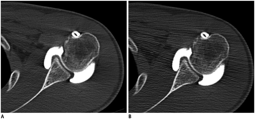
Fig. 1 Axial CT arthrographic images obtained by thin-slab-averaging reformation and conventional reconstruction in 24-year-old man for postoperative evaluation of SLAP lesion. A. Thin-slab-averaging reformation (used in this study). Image was obtained using two steps. Primary thin-section images were reconstructed using section thickness of 1.0 mm and reconstruction interval of 0.7 mm. Then, thin-slab-averaging reformation was applied to generate this image with section thickness of 2.0 mm and interval of 2.0 mm. B. Conventional reconstruction (for comparison). Image was reconstructed directly from raw projection data with section thickness of 2.0 mm and reconstruction interval of 2.0 mm. Image looks less smooth and noisier than that obtained by thin-slab-averaging reformation.
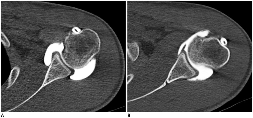
Fig. 2 Axial CT arthrographic images obtained at 120 kVp and 140 kVp in 24-year-old man for postoperative evaluation of superior labral anterior-to-posterior lesion. A. Image was taken in neutral position at 120 kVp, and shows better contrast between intraarticular contrast media solution and adjacent soft tissues and more noise than image obtained at 140 kVp. B. Image was taken in stress position at 140 kVp.
Reference
-
1. Lecouvet FE, Simoni P, Koutaïssoff S, Vande Berg BC, Malghem J, Dubuc JE. Multidetector spiral CT arthrography of the shoulder. Clinical applications and limits, with MR arthrography and arthroscopic correlations. Eur J Radiol. 2008; 68:120–136.2. Oh JH, Kim JY, Choi JA, Kim WS. Effectiveness of multidetector computed tomography arthrography for the diagnosis of shoulder pathology: comparison with magnetic resonance imaging with arthroscopic correlation. J Shoulder Elbow Surg. 2010; 19:14–20.3. Kim YJ, Choi JA, Oh JH, Hwang SI, Hong SH, Kang HS. Superior labral anteroposterior tears: accuracy and interobserver reliability of multidetector CT arthrography for diagnosis. Radiology. 2011; 260:207–215.4. De Filippo M, Araoz PA, Pogliacomi F, Castagna A, Petriccioli D, Sverzellati N, et al. Recurrent superior labral anterior-to-posterior tears after surgery: detection and grading with CT arthrography. Radiology. 2009; 252:781–788.5. Choi JY, Kim SH, Yoo HJ, Shin SH, Oh JH, Baek GH, et al. Superior labral anterior-to-posterior lesions: comparison of external rotation and active supination CT arthrography with neutral CT arthrography. Radiology. 2012; 263:199–205.6. Lecouvet FE, Dorzée B, Dubuc JE, Vande Berg BC, Jamart J, Malghem J. Cartilage lesions of the glenohumeral joint: diagnostic effectiveness of multidetector spiral CT arthrography and comparison with arthroscopy. Eur Radiol. 2007; 17:1763–1771.7. De Filippo M, Bertellini A, Sverzellati N, Pogliacomi F, Costantino C, Vitale M, et al. Multidetector computed tomography arthrography of the shoulder: diagnostic accuracy and indications. Acta Radiol. 2008; 49:540–549.8. Omoumi P, Bafort AC, Dubuc JE, Malghem J, Vande Berg BC, Lecouvet FE. Evaluation of rotator cuff tendon tears: comparison of multidetector CT arthrography and 1.5-T MR arthrography. Radiology. 2012; 264:812–882.9. Cochet H, Couderc S, Pelé E, Amoretti N, Moreau-Durieux MH, Hauger O. Rotator cuff tears: should abduction and external rotation (ABER) positioning be performed before image acquisition? A CT arthrography study. Eur Radiol. 2010; 20:1234–1241.10. Ambrosino MM, Genieser NB, Roche KJ, Kaul A, Lawrence RM. Feasibility of high-resolution, low-dose chest CT in evaluating the pediatric chest. Pediatr Radiol. 1994; 24:6–10.11. Sigal-Cinqualbre AB, Hennequin R, Abada HT, Chen X, Paul JF. Low-kilovoltage multi-detector row chest CT in adults: feasibility and effect on image quality and iodine dose. Radiology. 2004; 231:169–174.12. Heyer CM, Mohr PS, Lemburg SP, Peters SA, Nicolas V. Image quality and radiation exposure at pulmonary CT angiography with 100- or 120-kVp protocol: prospective randomized study. Radiology. 2007; 245:577–583.13. Wintersperger B, Jakobs T, Herzog P, Schaller S, Nikolaou K, Suess C, et al. Aorto-iliac multidetector-row CT angiography with low kV settings: improved vessel enhancement and simultaneous reduction of radiation dose. Eur Radiol. 2005; 15:334–341.14. Feuchtner GM, Jodocy D, Klauser A, Haberfellner B, Aglan I, Spoeck A, et al. Radiation dose reduction by using 100-kV tube voltage in cardiac 64-slice computed tomography: a comparative study. Eur J Radiol. 2010; 75:e51–e56.15. Park EA, Lee W, Kang JH, Yin YH, Chung JW, Park JH. The image quality and radiation dose of 100-kVp versus 120-kVp ECG-gated 16-slice CT coronary angiography. Korean J Radiol. 2009; 10:235–243.16. Abada HT, Larchez C, Daoud B, Sigal-Cinqualbre A, Paul JF. MDCT of the coronary arteries: feasibility of low-dose CT with ECG-pulsed tube current modulation to reduce radiation dose. AJR Am J Roentgenol. 2006; 186:6 Suppl 2. S387–S390.17. Choi JY, Kang HS, Hong SH, Lee JW, Kim NR, Jun WS, et al. Optimization of the contrast mixture ratio for simultaneous direct MR and CT arthrography: an in vitro study. Korean J Radiol. 2008; 9:520–525.18. Waldt S, Metz S, Burkart A, Mueller D, Bruegel M, Rummeny EJ, et al. Variants of the superior labrum and labro-bicipital complex: a comparative study of shoulder specimens using MR arthrography, multi-slice CT arthrography and anatomical dissection. Eur Radiol. 2006; 16:451–458.19. De Maeseneer M, Boulet C, Pouliart N, Kichouh M, Buls N, Verhelle F, et al. Assessment of the long head of the biceps tendon of the shoulder with 3T magnetic resonance arthrography and CT arthrography. Eur J Radiol. 2012; 81:934–939.20. Binkert CA, Verdun FR, Zanetti M, Pfirrmann CW, Hodler J. CT arthrography of the glenohumeral joint: CT fluoroscopy versus conventional CT and fluoroscopy--comparison of image-guidance techniques. Radiology. 2003; 229:153–158.21. Lee GY, Choi JA, Oh JH, Choi JY, Hong SH, Kang HS. Posteroinferior labral cleft at direct CT arthrography of the shoulder by using multidetector CT: is this a normal variant? Radiology. 2009; 253:765–770.22. von Falck C, Galanski M, Shin HO. Informatics in radiology: sliding-thin-slab averaging for improved depiction of low-contrast lesions with radiation dose savings at thin-section CT. Radiographics. 2010; 30:317–326.23. Joo SM, Lee KH, Kim YH, Kim SY, Kim K, Kim KJ, et al. Detection of the normal appendix with low-dose unenhanced CT: use of the sliding slab averaging technique. Radiology. 2009; 251:780–787.24. Lee KH, Hong H, Hahn S, Kim B, Kim KJ, Kim YH. Summation or axial slab average intensity projection of abdominal thin-section CT datasets: can they substitute for the primary reconstruction from raw projection data? J Digit Imaging. 2008; 21:422–432.
- Full Text Links
-
- Actions
-
Cited
- CITED
-
- Close
- Share
-
- Similar articles
-
- 100 kVp Low-Tube Voltage Abdominal CT in Adults: Radiation Dose Reduction and Image Quality Comparison of 120 kVp Abdominal CT
- Cervical Spine CT Using Spectral Shaping: Can It Be a Solution to Overcome Artifacts in the Lower Cervical Spinal Region?
- Windows Setting for Low kVp Abdominal CT: Comparison to 120-kVp CT Images
- Effects of Dual-Energy CT with Non-Linear Blending on Abdominal CT Angiography
- 80-kVp CT Using Iterative Reconstruction in Image Space Algorithm for the Detection of Hypervascular Hepatocellular Carcinoma: Phantom and Initial Clinical Experience
