Focal Anterior Displacement of the Thoracic Spinal Cord without Evidence of Spinal Cord Herniation or an Intradural Mass
- Affiliations
-
- 1Department of Radiology, Seoul National University Bundang Hospital, Seongnam 463-707, Korea. joonwoo2@gmail.com
- KMID: 1794644
- DOI: http://doi.org/10.3348/kjr.2014.15.6.733
Abstract
OBJECTIVE
We report magnetic resonance imaging (MRI) findings on focal anterior displacement of the thoracic spinal cord in asymptomatic patients without a spinal cord herniation or intradural mass.
MATERIALS AND METHODS
We identified 12 patients (male:female = 6:6; mean age, 51.7; range, 15-83 years) between 2007 and 2011, with focal anterior displacement of the spinal cord and without evidence of an intradural mass or spinal cord herniation. Two radiologists retrospectively reviewed the MRI findings in consensus.
RESULTS
An asymmetric spinal cord deformity with a focal dented appearance was seen on the posterior surface of the spinal cord in all patients, and it involved a length of 1 or 2 vertebral segments in the upper thoracic spine (thoracic vertebrae 1-6). Moreover, a focal widening of the posterior subarachnoid space was also observed in all cases. None of the patients had myelopathy symptoms, and they showed no focal T2-hyperintensity in the spinal cord with the exception of one patient. In addition, cerebrospinal fluid (CSF) flow artifacts were seen in the posterior subarachnoid space of the affected spinal cord level. Computed tomography myelography revealed preserved CSF flow in the two available patients.
CONCLUSION
Focal anterior spinal cord indentation can be found in the upper thoracic level of asymptomatic patients without a spinal cord herniation or intradural mass.
Keyword
MeSH Terms
-
Adolescent
Adult
Aged
Aged, 80 and over
Cerebrospinal Fluid/physiology
Female
Hernia/pathology
Humans
Magnetic Resonance Imaging
Male
Middle Aged
Retrospective Studies
Spinal Cord Diseases/pathology/*radiography/surgery
Spine/pathology/radiography
Thoracic Vertebrae/pathology/*radiography
Tomography, X-Ray Computed
Young Adult
Figure
-

Fig. 1 Whole-spine sagittal T2-weighted images show incidental findings of focal anterior displacement of thoracic spinal cord in two patients. A, B. Asymmetric dented appearance with ventral angulation of cord and widening of dorsal cerebrospinal fluid (CSF) space are present without intradural mass or spinal cord herniation in upper thoracic spine. Prominent CSF flow artifacts are present in posterior subarachnoid space at involved spine level. Arrow indicates spinal cord.

Fig. 2 Cervical spine magnetic resonance imaging (A-D) and additional computed tomography (CT) myelography (E, F) of 38-year-old woman who presented with neck pain. Sagittal T2-weighted image (A) and axial T2-weighted image (B) show anteriorly displaced spinal cord at T2/3 level with hypointense elongated flow artifact in extended dorsal space at same level. Sagittal (C) and axial (D) CT myelography in supine position show widening of posterior space without intradural filling defect or blockade (such as those caused by arachnoid cyst). Sagittal (E) and axial (F) CT myelography in prone position show preservation of anterior subarachnoid space and intact cerebrospinal fluid flow. There was no finding of spinal cord herniation or spinal cord adhesion. All axial images (B, D, F) were obtained at T2 level. Arrow indicates spinal cord.
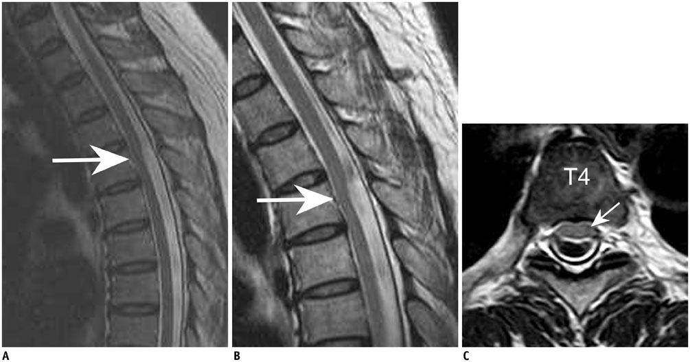
Fig. 3 Lumbosacral spine magnetic resonance imaging (MRI) of 48-year-old woman who presented with low back pain. A. Sagittal T2-weighted image shows asymmetric dented appearance and anteriorly displaced spinal cord at T4 level. B, C. Follow-up cervical spine MRI study 4 months after first examination. Sagittal T2-weighted image (B) shows no interval change in cord deformity. Axial T2-weighted image (C) shows ventral displacement of cord without focal spinal cord herniation and relatively preserved anterior subarachnoid space at T4 level. Arrow indicates spinal cord.
Reference
-
1. Parmar H, Park P, Brahma B, Gandhi D. Imaging of idiopathic spinal cord herniation. Radiographics. 2008; 28:511–518.2. Groen RJ, Coppes MH. Operative treatment of posterior spinal arachnoid cysts: do not refrain from checking on an anterior transdural spinal cord herniation. Acta Neurochir (Wien). 2011; 153:601–602. author reply 603.3. van den Hauwe L, Van Goethem JW, Goedseels K, Merlevede K, Degryse H, Parizel PM. Thoracic spinal cord herniation and arachnoid cyst. JBR-BTR. 2006; 89:150–115.4. Dix JE, Griffitt W, Yates C, Johnson B. Spontaneous thoracic spinal cord herniation through an anterior dural defect. AJNR Am J Neuroradiol. 1998; 19:1345–1348.5. Reardon MA, Raghavan P, Carpenter-Bailey K, Mukherjee S, Smith JS, Matsumoto JA, et al. Dorsal thoracic arachnoid web and the "scalpel sign": a distinct clinical-radiologic entity. AJNR Am J Neuroradiol. 2013; 34:1104–1110.6. Brus-Ramer M, Dillon WP. Idiopathic thoracic spinal cord herniation: retrospective analysis supporting a mechanism of diskogenic dural injury and subsequent tamponade. AJNR Am J Neuroradiol. 2012; 33:52–56.7. Tekkök IH. Spontaneous spinal cord herniation: case report and review of the literature. Neurosurgery. 2000; 46:485–491. discussion 491-492.8. Sasani M, Ozer AF, Vural M, Sarioglu AC. Idiopathic spinal cord herniation: case report and review of the literature. J Spinal Cord Med. 2009; 32:86–94.9. Groen RJ, Middel B, Meilof JF, de Vos-van de Biezenbos JB, Enting RH, Coppes MH, et al. Operative treatment of anterior thoracic spinal cord herniation: three new cases and an individual patient data meta-analysis of 126 case reports. Neurosurgery. 2009; 64:3 Suppl. ons145–ons159. discussion ons159-ons160.10. Taylor TR, Dineen R, White B, Jaspan T. The thoracic anterior spinal cord adhesion syndrome. Br J Radiol. 2012; 85:e123–e129.11. Abou-Fakhr FS, Kanaan SV, Youness FM, Hourani MH, Haddad MC. Thoracic spinal intradural arachnoid cyst: report of two cases and review of literature. Eur Radiol. 2002; 12:877–882.12. Kumar K, Malik S, Schulte PA. Symptomatic spinal arachnoid cysts: report of two cases with review of the literature. Spine (Phila Pa 1976). 2003; 28:E25–E29.13. Wang MY, Levi AD, Green BA. Intradural spinal arachnoid cysts in adults. Surg Neurol. 2003; 60:49–55. discussion 55-56.14. Najjar MW, Baeesa SS, Lingawi SS. Idiopathic spinal cord herniation: a new theory of pathogenesis. Surg Neurol. 2004; 62:161–170. discussion 170-171.
- Full Text Links
-
- Actions
-
Cited
- CITED
-
- Close
- Share
-
- Similar articles
-
- Spontaneous Herniation of the Thoracic Spinal Cord: A Case Report
- Unilateral Paramedian Transpedicular Approach for Repair of Anterior Transdural Spinal Cord Herniation: Report of a Case and Literature Review
- Intradural Lipoma in the Lower Thoracic Spinal Cord
- A Case of Intradural Spinal Lipoma
- Spontaneous Anterior Thoracic Spinal Cord Herniation through Dura Defect: A Case Report
