Contrast-Enhanced Spectral Mammography: Comparison with Conventional Mammography and Histopathology in 152 Women
- Affiliations
-
- 1Department of Radiology, Centre of Oncology, Maria Sklodowska-Curie Memorial Institute, Krakow 31-115, Poland.
- 2Department of Gynecologic Oncology, Centre of Oncology, Maria Sklodowska-Curie Memorial Institute, Krakow 31-115, Poland. pawel.blecharz@interia.pl
- 3Department of Tumour Pathology, Centre of Oncology, Maria Sklodowska-Curie Memorial Institute, Krakow 31-115, Poland.
- 4Department of Radiotherapy, Centre of Oncology, Maria Sklodowska-Curie Memorial Institute, Krakow 31-115, Poland.
- KMID: 1794639
- DOI: http://doi.org/10.3348/kjr.2014.15.6.689
Abstract
OBJECTIVE
The goal of the study was to compare conventional mammography (MG) and contrast-enhanced spectral mammography (CESM) in preoperative women.
MATERIALS AND METHODS
The study was approved by the local Ethics Committee and all participants provided informed consent. The study included 152 consecutive patients with 173 breast lesions diagnosed on MG or CESM. All MG examinations and consults were conducted in one oncology centre. Non-ionic contrast agent, at a total dose of 1.5 mL/kg body weight, was injected intravenous. Subsequently, CESM exams were performed with a mammography device, allowing dual-energy acquisitions. The entire procedure was done within the oncology centre. Images from low and high energy exposures were processed together and the combination provided an "iodine" image which outlined contrast up-take in the breast.
RESULTS
MG detected 157 lesions in 150 patients, including 92 infiltrating cancers, 12 non-infiltrating cancers, and 53 benign lesions. CESM detected 149 lesions in 128 patients, including 101 infiltrating cancers, 13 non-infiltrating cancers, and 35 benign lesions. CESM sensitivity was 100% (vs. 91% for MG), specificity was 41% (vs. 15% for MG), area under the receiver operating characteristic curve was 0.86 (vs. 0.67 for MG), and accuracy was 80% (vs. 65% for MG) for the diagnosis of breast cancer. Both MG and CESM overestimated lesion sizes compared to histopathology (p < 0.001).
CONCLUSION
CESM may provide higher sensitivity for breast cancer detection and greater diagnostic accuracy than conventional mammography.
MeSH Terms
Figure
-
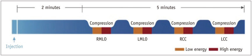
Fig. 1 Contrast-enhanced spectral mammography examination scheme (example where left breast is most suspicious) (Courtesy of GE Healthcare). LCC = left craniocaudal, LMLO = left mediolateral oblique, RCC = right craniocaudal, RMLO = right mediolateral oblique
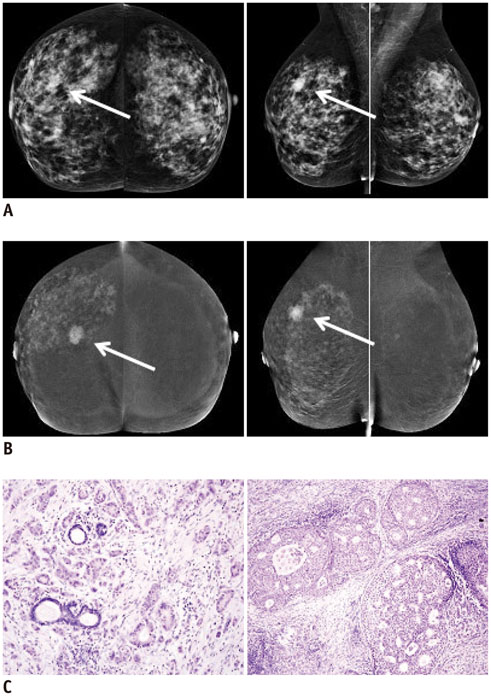
Fig. 2 Infiltrating ductal carcinoma (arrows) of right breast in 63-year-old woman with dense fibroglandular breast tissue. A. Digital conventional mammography (MG) with mediolateral oblique (MLO) view of right breast reveals oval, well demarcated shadow 15 mm in diameter. This mass is not visible in MG craniocaudal (CC) projection. B. Contrast-enhanced spectral mammography in same patient shows poorly demarcated focus of enhancement 15 mm in diameter visible in both MLO and CC projections. Additionally, in right breast, small enhancing foci are visible in upper and lower outer quadrants. C. In histopathological examination, infiltrating carcinoma (of no special type) grade 2 was found within main focus of enhancement. Ductal carcinoma in situ (DCIS) component was noticed in unaltered areas macroscopically. Hematoxylin and eosin staining. Objective magnification 10 × (invasive component, left) and 20 × (DCIS, right).
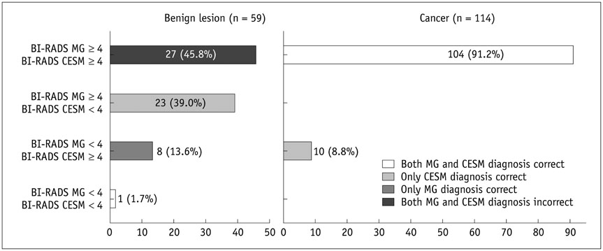
Fig. 3 Comparison of correct and incorrect assessments on conventional mammography (MG) and contrast-enhanced spectral mammography (CESM) based on Breast Imaging Reporting and Data System (BI-RADS) scores (BI-RADS scores on MG and CESM were consistent and correct in 91% of proven malignant lesions). Diagnosis was correct if lesion scored as BI-RADS < 4 was benign or scored ≥ 4 was malignant. Left picture presents agreement between MG and CESM diagnoses with pathological results in 59 benign lesions, right picture-in 114 malignant lesions.
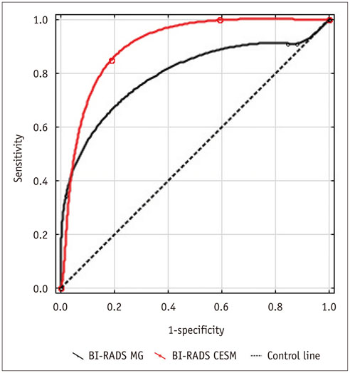
Fig. 4 Receiver operating characteristic curves from Breast Imaging Reporting and Data System (BI-RADS) conventional mammography (MG) and contrast-enhanced spectral mammography (CESM) assessments.
Reference
-
1. Jochelson M. Advanced imaging techniques for the detection of breast cancer. Am Soc Clin Oncol Educ Book. 2012; 65–69.2. Burhenne HJ, Burhenne LW, Goldberg F, Hislop TG, Worth AJ, Rebbeck PM, et al. Interval breast cancers in the Screening Mammography Program of British Columbia: analysis and classification. AJR Am J Roentgenol. 1994; 162:1067–1071. discussion 1072-1075.3. Robertson CL. A private breast imaging practice: medical audit of 25,788 screening and 1,077 diagnostic examinations. Radiology. 1993; 187:75–79.4. Mandelson MT, Oestreicher N, Porter PL, White D, Finder CA, Taplin SH, et al. Breast density as a predictor of mammographic detection: comparison of interval- and screen-detected cancers. J Natl Cancer Inst. 2000; 92:1081–1087.5. Kolb TM, Lichy J, Newhouse JH. Comparison of the performance of screening mammography, physical examination, and breast US and evaluation of factors that influence them: an analysis of 27,825 patient evaluations. Radiology. 2002; 225:165–175.6. Pisano ED, Hendrick RE, Yaffe MJ, Baum JK, Acharyya S, Cormack JB, et al. Diagnostic accuracy of digital versus film mammography: exploratory analysis of selected population subgroups in DMIST. Radiology. 2008; 246:376–383.7. Pisano ED, Gatsonis C, Hendrick E, Yaffe M, Baum JK, Acharyya S, et al. Diagnostic performance of digital versus film mammography for breast-cancer screening. N Engl J Med. 2005; 353:1773–1783.8. Fischer U, Baum F, Obenauer S, Luftner-Nagel S, von Heyden D, Vosshenrich R, et al. Comparative study in patients with microcalcifications: full-field digital mammography vs screen-film mammography. Eur Radiol. 2002; 12:2679–2683.9. Smith A. Fundamentals of digital mammography: physics, technology and practical considerations. Radiol Manage. 2003; 25:18–24. 26–31. quiz 32-34.10. Knopp MV, Giesel FL, Marcos H, von Tengg-Kobligk H, Choyke P. Dynamic contrast-enhanced magnetic resonance imaging in oncology. Top Magn Reson Imaging. 2001; 12:301–308.11. Padhani AR, Dzik-Jurasz A. Perfusion MR imaging of extracranial tumor angiogenesis. Top Magn Reson Imaging. 2004; 15:41–57.12. Jeswani T, Padhani AR. Imaging tumour angiogenesis. Cancer Imaging. 2005; 5:131–138.13. Schäfer AO, Langer M. [MRI mammography screening in women with lobular carcinoma in situ]. Strahlenther Onkol. 2012; 188:716–717.14. Jochelson MS, Dershaw DD, Sung JS, Heerdt AS, Thornton C, Moskowitz CS, et al. Bilateral contrast-enhanced dual-energy digital mammography: feasibility and comparison with conventional digital mammography and MR imaging in women with known breast carcinoma. Radiology. 2013; 266:743–751.15. Berg WA. Rationale for a trial of screening breast ultrasound: American College of Radiology Imaging Network (ACRIN) 6666. AJR Am J Roentgenol. 2003; 180:1225–1228.16. Dromain C, Balleyguier C, Muller S, Mathieu MC, Rochard F, Opolon P, et al. Evaluation of tumor angiogenesis of breast carcinoma using contrast-enhanced digital mammography. AJR Am J Roentgenol. 2006; 187:W528–W537.17. Dromain C, Thibault F, Diekmann F, Fallenberg EM, Jong RA, Koomen M, et al. Dual-energy contrast-enhanced digital mammography: initial clinical results of a multireader, multicase study. Breast Cancer Res. 2012; 14:R94.18. Puong S, Bouchevreau X, Patoureaux F, Iordache R, Muller S. Dual-energy contrast enhanced digital mammography using a new approach for breast tissue canceling. Proc SPIE. 2007; 6510:65102H.19. Diekmann F, Freyer M, Diekmann S, Fallenberg EM, Fischer T, Bick U, et al. Evaluation of contrast-enhanced digital mammography. Eur J Radiol. 2011; 78:112–121.20. Boetes C, Mus RD, Holland R, Barentsz JO, Strijk SP, Wobbes T, et al. Breast tumors: comparative accuracy of MR imaging relative to mammography and US for demonstrating extent. Radiology. 1995; 197:743–747.21. Orel SG, Schnall MD. MR imaging of the breast for the detection, diagnosis, and staging of breast cancer. Radiology. 2001; 220:13–30.22. Kerlikowske K, Grady D, Barclay J, Sickles EA, Ernster V. Effect of age, breast density, and family history on the sensitivity of first screening mammography. JAMA. 1996; 276:33–38.23. Lewin JM, Niklason L. Advanced applications of digital mammography: tomosynthesis and contrast-enhanced digital mammography. Semin Roentgenol. 2007; 42:243–252.
- Full Text Links
-
- Actions
-
Cited
- CITED
-
- Close
- Share
-
- Similar articles
-
- Digital Mammography
- Contrast-Enhanced Spectral Mammography Versus Ultrasonography: Diagnostic Performance in Symptomatic Patients with Dense Breasts
- Decisional balance corresponding to the Stage of Adoption for Mammography in Middle Aged Women
- Comparison of Breast Cancer Screening Results in Korean Middle-Aged Women: A Hospital-based Prospective Cohort Study
- Contrast-Enhanced Spectral Mammography: Importance of the Assessment of Breast Tumor Size
