Isolated Colonic Schwannoma in the Ascending Colon: A Case Report and Literature Review of Schwannomas in the Large Intestine
- Affiliations
-
- 1Department of Radiology, Haeundae Paik Hospital, Inje University College of Medicine, Busan, Korea. chosai81@gmail.com
- 2Department of Pathology, Haeundae Paik Hospital, Inje University College of Medicine, Busan, Korea.
- KMID: 1793894
- DOI: http://doi.org/10.3348/jksr.2015.72.5.358
Abstract
- Schwannomas are benign mesenchymal spindle cell tumors arising from the Schwann cells that form the peripheral neural sheath. Several recent studies indicate that although reports of gastrointestinal schwannomas have increased with advanced technological developments in immunohistochemical staining, isolated colonic schwannomas are extremely rare. Moreover, it is known to be somewhat difficult to diagnose colonic schwannoma before surgical operation. In this paper, we report a case of isolated schwannoma that was incidentally discovered in the ascending colon, along with a review of few recent literatures.
Figure
-
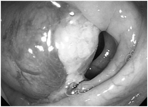
Fig. 1 A colonoscopy reveals an intraluminal round submucosal tumor, which is measured approximately 4 cm in diameter, in the ascending colon.
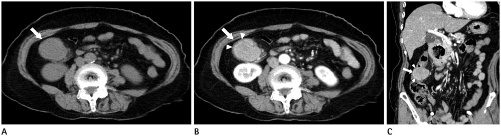
Fig. 2 Pre-contrast (A) and contrast enhanced axial (B) and coronal (C) CT scan demonstrates a lobulating contoured mass (arrow) in the ascending colon. On pre-contrast axial CT scan (A), there is no calcification or cystic component. Enhanced axial (B) and coronal reformated (C) CT images show homogeneous enhancement of the mass and intact overlying mucosa (arrowheads).

Fig. 3 Gross examination of the ascending colon revealed a yellowish gray fungating mass measuring 4.1 × 3.4 cm.
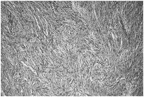
Fig. 4 Photomicrography demonstrates proliferation of plump spindle cells with occasional fascicular pattern, lacking significant nuclear atypia (hematoxylin and eosin, × 100).
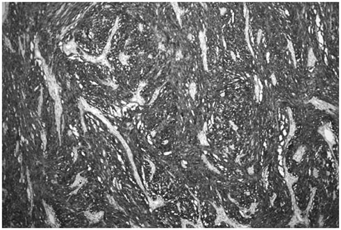
Fig. 5 Immunohistochemical staining for S-100 protein is diffuse and strong (× 100).
Reference
-
1. Levy AD, Quiles AM, Miettinen M, Sobin LH. Gastrointestinal schwannomas: CT features with clinicopathologic correlation. AJR Am J Roentgenol. 2005; 184:797–802.2. Inagawa S, Hori M, Shimazaki J, Matsumoto S, Ishii H, Itabashi M, et al. Solitary schwannoma of the colon: report of two cases. Surg Today. 2001; 31:833–838.3. Hou YY, Tan YS, Xu JF, Wang XN, Lu SH, Ji Y, et al. Schwannoma of the gastrointestinal tract: a clinicopathological, immunohistochemical and ultrastructural study of 33 cases. Histopathology. 2006; 48:536–545.4. Prévot S, Bienvenu L, Vaillant JC, de Saint-Maur PP. Benign schwannoma of the digestive tract: a clinicopathologic and immunohistochemical study of five cases, including a case of esophageal tumor. Am J Surg Pathol. 1999; 23:431–436.5. Kanneganti K, Patel H, Niazi M, Kumbum K, Balar B. Cecal schwannoma: a rare cause of gastrointestinal bleeding in a young woman with review of literature. Gastroenterol Res Pract. 2011; 2011:142781.6. Tomozawa S, Masaki T, Matsuda K, Yokoyama T, Ishida T, Muto T. A schwannoma of the cecum: case report and review of Japanese schwannomas in the large intestine. J Gastroenterol. 1998; 33:872–875.7. Choi JW, Choi D, Kim KM, Sohn TS, Lee JH, Kim HJ, et al. Small submucosal tumors of the stomach: differentiation of gastric schwannoma from gastrointestinal stromal tumor with CT. Korean J Radiol. 2012; 13:425–433.8. Lee SH, Kim TO, Hwang SY, Ryu DY, Lee DH, Park WI, et al. [A case of rectal schwannoma presenting with hematochezia]. Korean J Gastroenterol. 2006; 48:195–199.9. Kang DB, Kim SH, Oh JT, Park WC, Lee JK, Kim HS. Colonic schwannoma. J Korean Surg Soc. 2007; 73:183–187.10. Min YW, Kim YH, Yun HS, Kil JS, Kim YC, Yun SH, et al. [A case of benign schwannoma in the ascending colon]. Korean J Gastroenterol. 2007; 50:398–401.11. Tsunoda C, Kato H, Sakamoto T, Yamada R, Mitsumaru A, Yokomizo H, et al. A case of benign schwannoma of the transverse colon with granulation tissue. Case Rep Gastroenterol. 2009; 3:116–120.12. Hsu W, Wu I, Chen C, Liu C, Chen H, Wu D. Colon schwannoma: a case report. J Intern Med Taiwan. 2009; 20:255–259.13. Park KJ, Kim KH, Roh YH, Kim SH, Lee J, Rha S, et al. Isolated primary schwannoma arising on the colon: report of two cases and review of the literature. J Korean Surg Soc. 2011; 80:367–372.14. Matsumoto T, Yamamoto S, Fujita S, Akasu T, Moriya Y. Cecal schwannoma with laparoscopic wedge resection: report of a case. Asian J Endosc Surg. 2011; 4:178–180.15. Vasilakaki T, Skafida E, Arkoumani E, Grammatoglou X, Tsavari KK, Myoteri D, et al. Synchronous primary adenocarcinoma and ancient schwannoma in the colon: an unusual case report. Case Rep Oncol. 2012; 5:164–168.16. Kim HJ, Kim CH, Lim SW, Huh JW, Kim YJ, Kim HR. Schwannoma of ascending colon treated by laparoscopic right hemicolectomy. World J Surg Oncol. 2012; 10:81.17. Baek SJ, Hwangbo W, Kim J, Kim IS. A case of benign schwannoma of the ascending colon treated with laparoscopic-assisted wedge resection. Int Surg. 2013; 98:315–318.18. de Mesquita Neto JW, Lima Verde Leal RM, de Brito EV, Cordeiro DF, Costa ML. Solitary schwannoma of the cecum: case report and review of the literature. Case Rep Oncol. 2013; 6:62–65.19. Jung EJ, Han HS, Koh YC, Cho J, Ryu CG, Paik JH, et al. Metachronous schwannoma in the colon with vestibular schwannoma. Ann Surg Treat Res. 2014; 87:161–165.
- Full Text Links
-
- Actions
-
Cited
- CITED
-
- Close
- Share
-
- Similar articles
-
- A Case of Benign Schwannoma in the Ascending Colon
- Isolated primary schwannoma arising on the colon: report of two cases and review of the literature
- A Case Report: Schwannoma of the Auricle
- Surgical treatment of multiple plexiform schwannomas arising from the superficial radial nerve: a case report
- A Case of Lymphangiomatosis Arising in the Colon
