Surgical treatment of multiple plexiform schwannomas arising from the superficial radial nerve: a case report
- Affiliations
-
- 1Department of Orthopaedic Surgery, Pohang St. Mary’s Hospital, Pohang, Korea
- KMID: 2536227
- DOI: http://doi.org/10.12790/ahm.22.0031
Abstract
- Schwannoma, or neurilemmoma, is a benign neoplasm that arises from Schwann cells, which surround peripheral, cranial, and autonomic nerve sheaths. Schwannoma has been reported to occur mainly as a singular lesion of the sacral nerve or sciatic nerve in young adults. Plexiform schwannoma, a subtype of schwannoma, is a rare neoplasm known to account for 2% to 5% of total schwannomas. Schwannoma of the upper extremities is relatively rare and is reported to occur mostly in the ulnar nerve. We report, with a literature review, a case of 4.2-cm and 2.8-cm symptomatic multiple plexiform schwannomas that occurred in the superficial radial nerve and were treated without neurologic sequelae by surgical resection.
Keyword
Figure
-

Fig. 1. Preoperative clinical photographs of the patient. Multiple masses presented in the anterolateral aspect of patients left forearm. (A) In the anterior view, each sized 5 cm and 3.5 cm, circular masses were located in the anterior side of proximal and distal area of forearm. (B) In the lateral view, large size mass was located in the proximal area of forearm.
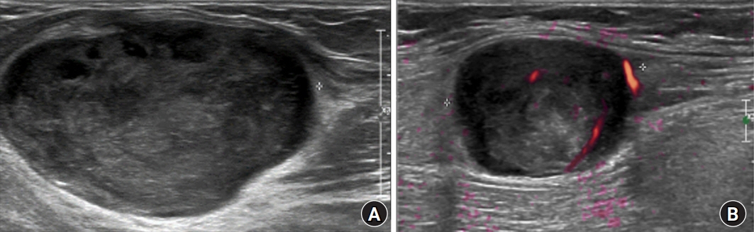
Fig. 2. Ultrasound images. (A) An ovoid, hypoechoic lesion with a length of 4.2 cm was observed in the anterior aspect of the left elbow, connected to the superficial radial nerve on an ultrasound examination. (B) An ovoid, hypoechoic lesion with a length of 2.8 cm was observed in the anterior aspect of the left forearm, located near the median nerve, but the connection was uncertain based on the ultrasound examination.
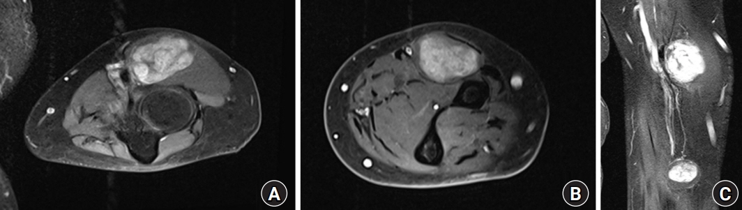
Fig. 3. Magnetic resonance images at the level of the forearm demonstrate areas of low signal intensity on T1-weighted imaging and intermediate to high signal intensity on T2-weighted imaging that are connected to the superficial radial nerve. (A) A mass with a length of 4.5 cm is observed on the anterior aspect of the elbow. (B) A mass with a length of 3.0 cm is observed on the anterior aspect of the forearm. (C) Two masses are observed on the sagittal view.
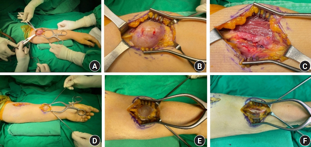
Fig. 4. Intraoperative clinical photographs of multiple schwannomas of the upper arm at the level of the elbow (A–C) and the anterior aspect of the mid-forearm (D–F).
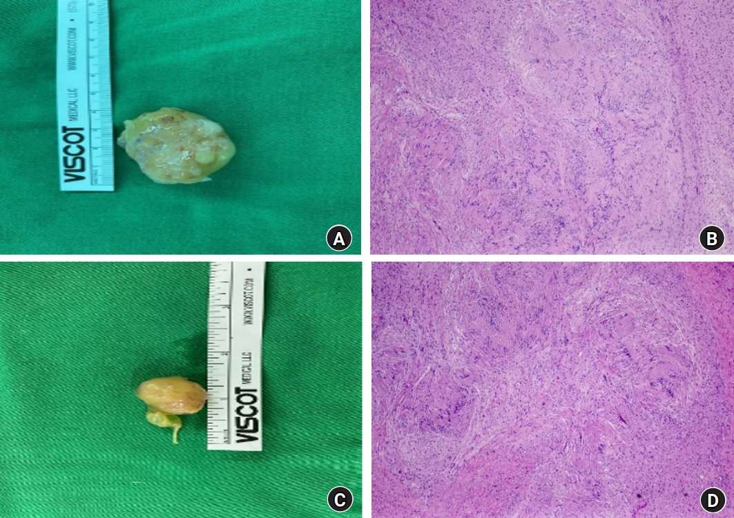
Fig. 5. Light microscopic findings and gross mass images of the left elbow (A, B) and forearm (C, D). The tumors are cases of plexiform neurilemmomatosis, showing multilobulated nodules with hypercellular Antoni A areas and hypocellular Antoni B areas (hematoxylin and eosin stain, ×40).

Fig. 6. Well-defined multinodular and biphasic tumor (Masson’s trichrome). (A) ×15, (B) ×40.
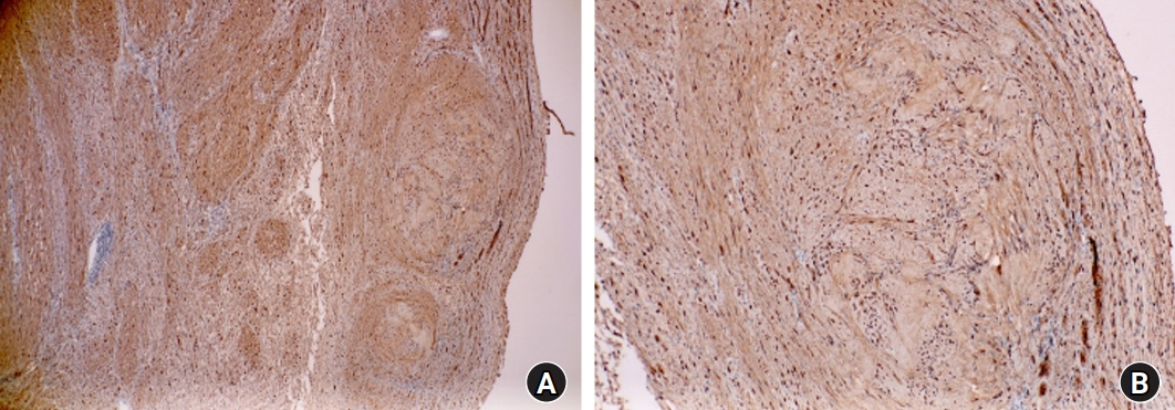
Fig. 7. Immunohistochemical staining of tumor nodules shows strong positivity for S-100 protein (×40). (A) Mass on the anterior aspect of the elbow. (B) Mass on the anterior aspect of the forearm.
Reference
-
References
1. Li XN, Cui JL, Christopasak SP, Kumar A, Peng ZG. Multiple plexiform schwannomas in the plantar aspect of the foot: case report and literature review. BMC Musculoskelet Disord. 2014; 15:342.
Article2. Enziger FM, Weiss SW. Soft tissue tumors. 3rd ed. St. Louis: Mosby;1995. p. 821–88.3. Perrotta R, Virzì D, Tarico MS, Napoli P. An unusual case of symptomatic schwannoma on the elbow. Br J Neurosurg. 2011; 25:306–7.
Article4. Kransdorf MJ. Benign soft-tissue tumors in a large referral population: distribution of specific diagnoses by age, sex, and location. AJR Am J Roentgenol. 1995; 164:395–402.
Article5. Harkin JH, Arrington JH, Reed RJ. Benign plexiform schwannoma, a lesion distinct from plexiform neurofibroma. J Neuropathol Exp Neurol. 1978; 37:622.
Article6. Daras M, Koppel BS, Heise CW, Mazzeo MJ, Poon TP, Duffy KR. Multiple spinal intradural schwannomas in the absence of von Recklinghausen’s disease. Spine (Phila Pa 1976). 1993; 18:2556–9.
Article7. Cerofolini E, Landi A, DeSantis G, Maiorana A, Canossi G, Romagnoli R. MR of benign peripheral nerve sheath tumors. J Comput Assist Tomogr. 1991; 15:593–7.
Article8. Stull MA, Moser RP Jr, Kransdorf MJ, Bogumill GP, Nelson MC. Magnetic resonance appearance of peripheral nerve sheath tumors. Skeletal Radiol. 1991; 20:9–14.
Article
