A Case of Early Bile Duct Cancer Arising from Villous Adenoma in Choledochal Cyst
- Affiliations
-
- 1Department of Internal Medicine, KwangMyung SungAe Hospital, Gwangmyeong, Korea. kimhkgi@naver.com
- 2Department of Internal Medicine, SungAe Hospital, Seoul, Korea.
- 3Department of Radiology, KwangMyung SungAe Hospital, Gwangmyeong, Korea.
- KMID: 1792768
- DOI: http://doi.org/10.4166/kjg.2009.54.1.55
Abstract
- Choledochal cyst is an uncommon premalignant anomaly. The morphology and pathogenesis of the premalignant lesion of cholangiocarcinoma arising from the choledochal cyst has not been well described. Herein, we report a rare case of bile duct adenoma arising from choledochal cyst with anomalous union of pancreaticobiliary duct (AUPBD). 50-year-old woman was admitted to our hospital with the complaint of epigastric pain. She had received common bile duct (CBD) exploration and choledocholithotomy and cholecystectomy 3 months earlier under the diagnosis of multiple CBD stones. Intraoperalive cholangiogram was not remarkable except CBD dilatation at that time. Endoscopic retrograde cholangiopancreatography revealed choledochal cyst with AUPBD and round filling defect which disappeared easily on the balloon cholaniogram. On magnetic resonance cholangiopancreatography, the filling defect was confirmed as 2 cm polypoid mass attached to the distal bile duct wall. At laparotomy, a soft whitish mass was palpable on the lower CBD. On histological examination, adenoma with focal carcinoma change arising from choledochal cyst was diagnosed.
MeSH Terms
Figure
-
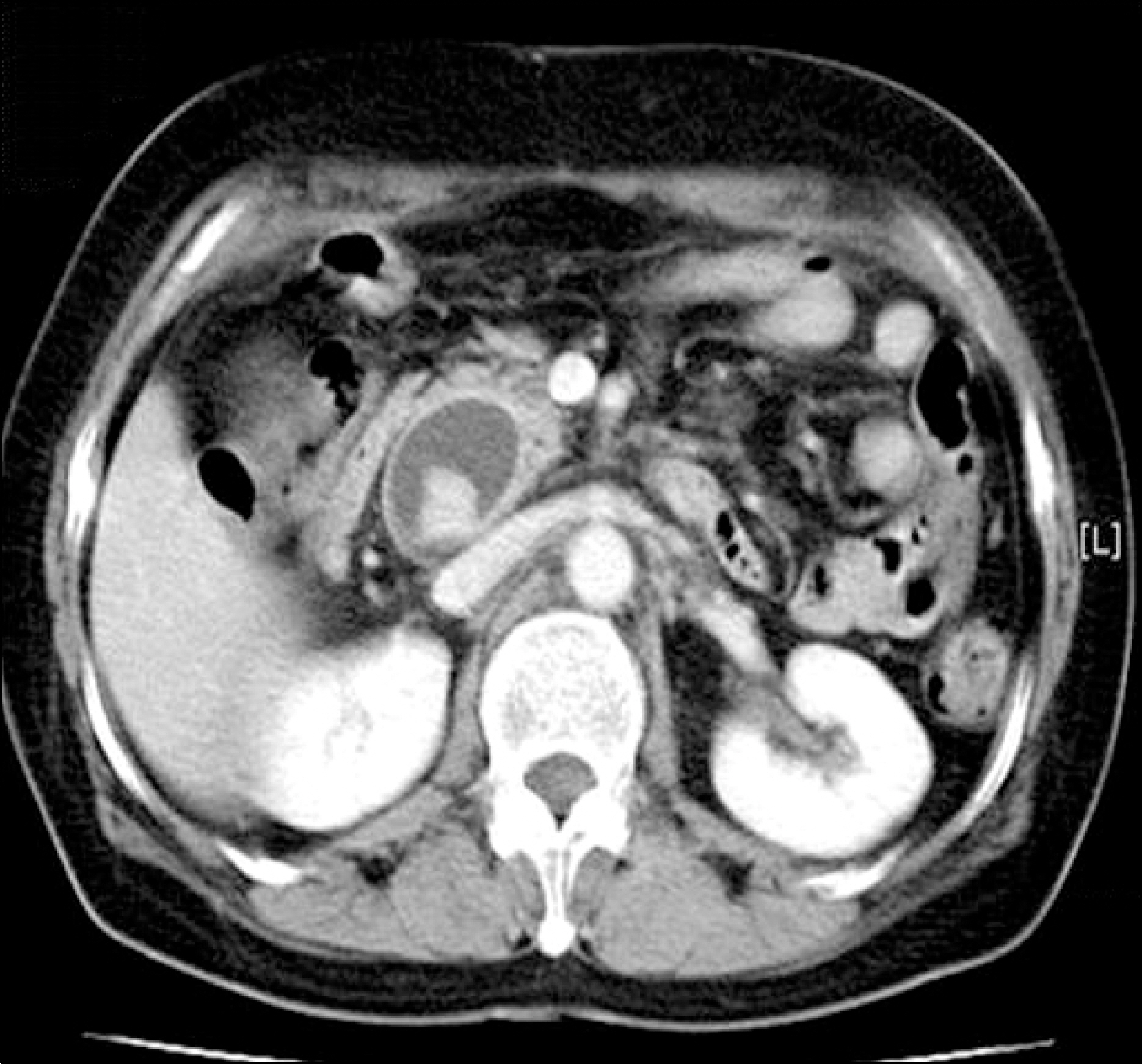
Fig. 1. Abdominal CT showed multiple bile duct stones with marked common bile duct dilatation.
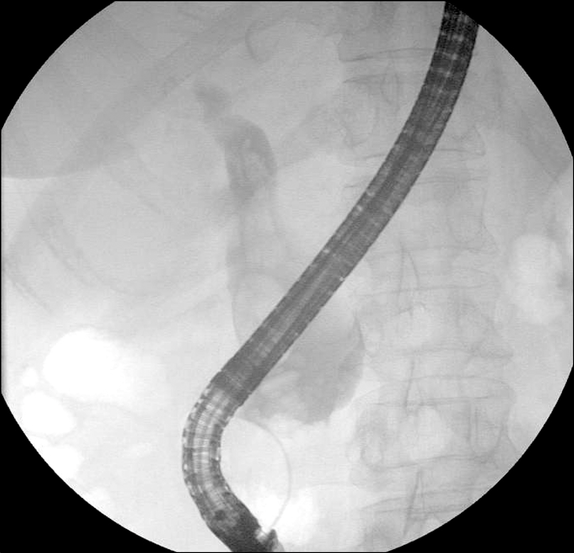
Fig. 2. ERCP shows multiple filling defect of common bile duct and proximal duct dilatation. But, basket extraction and balloon sweeping retrieved nothing. Pancreatic duct was not cannulated.

Fig. 3. ERCP showed round filling defect which disappeared easily on the balloon occlusive cholangiogram (A) and revealed choledochal cyst with anomalous union of pancreaticobiliary duct (AUPBD) (B).
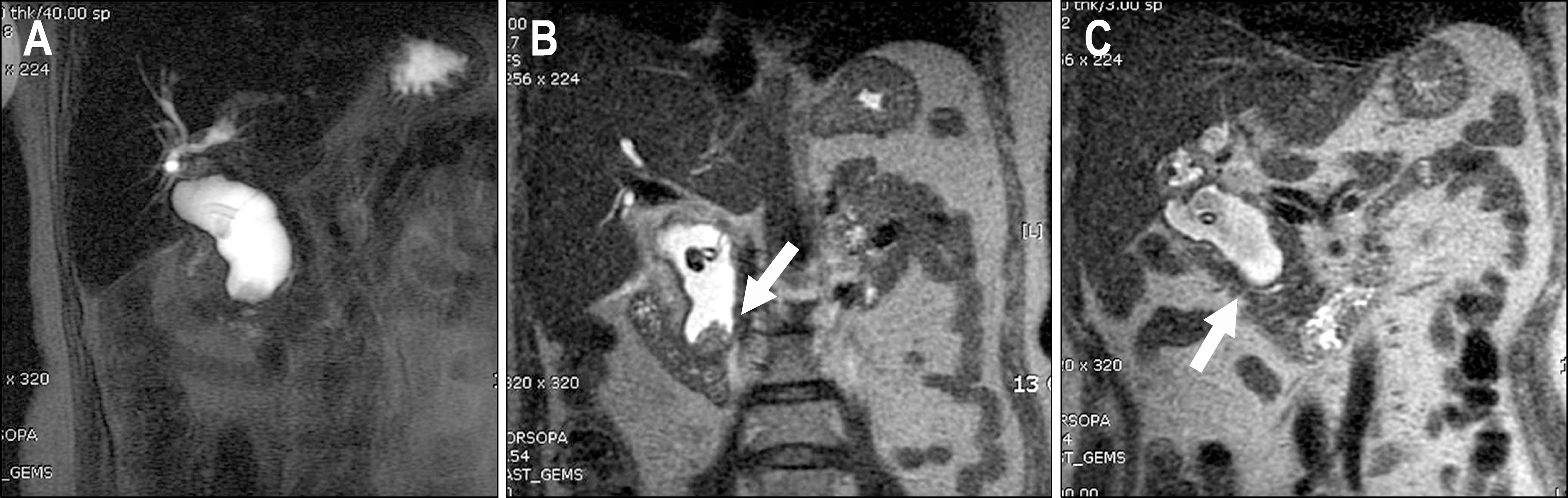
Fig. 4. MRCP showed fusiform dilatation of extrahepatic duct (A) and a soft tissue signal mass measured 2×2 cm within a choledochal cyst (B). Combined AUPBD with choledochal cyst was seen (C).
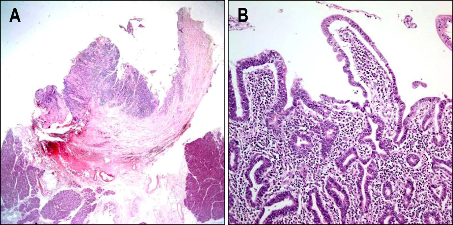
Fig. 5. (A) Microscopic findings of the specimen showed protruding polypoid lesion into lumen, consisting of tubulovillous adenoma (H&E stain, ×20). (B) Focal adenocarcinoma change arising from adenoma was lim-ited to bile duct mucosa (H&E stain, ×200).
Reference
-
1. Kim MH, Yoo BM, Seo DW, et al. Choledochal cyst and anomalous union of pancreaticobiliary duct in the adult. Korean J Gastrointest Endosc. 1996; 16:41–48.2. Lipsett PA, Locke JE. Biliary cystic disease. Curr Treat Options Gastroenterol. 2006; 9:107–112.
Article3. Franko J, Nussbaum ML, Morris JB. Choledochal cyst cholangiocarcinoma arising from adenoma: case report and a review of the literature. Curr Surg. 2006; 63:281–284.
Article4. Komi N, Tamura T, Tsuge S, Miyoshi Y, Udaka H, Takehara H. Relation of patient age to premalignant alterations in choledochal cyst epithelium: histochemical and immunohistochemical studies. J Pediatr Surg. 1986; 21:430–433.
Article5. Nagata E, Sakai K, Kinoshata H, Hirohashi K. Choledochal cyst: complications of anomalous connection between the choledochus and pancreatic and carcinoma of the biliary tract. World J Surg. 1986; 10:102–110.6. Aggarwal S, Kumar S, Kumar A, Bhasin R, Garg PK, Bandhu S. Extrahepatic bile duct adenoma in a patient with a choledochal cyst. J Gastroenterol Hepatol. 2003; 18:351–352.
Article7. Kawakami H, Kuwatani M, Onodera M, Asaka M, Hirano S, Kondo S. Villous adenoma arising in choledochocele. Gastrointest Endosc. 2007; 66:1231–1232.
Article8. Kim JS, Lee SJ, Yeon JE, et al. A case of adenoma of the common bile duct originating at the cystic duct opening. Korean J Gastrointest Endosc. 1995; 15:91–97.9. Imazu M, Iwai N, Tokiwa K, Shimotake T, Kimura O, Ono S. Factors of biliary carcinogenesis in choledochal cysts. Eur J Pediatr Surg. 2001; 11:24–27.
Article10. Metcalfe MS, Wemyss-Holden SA, Maddern GJ. Management dilemmas with choledochal cysts. Arch Surg. 2003; 138:333–339.
Article11. Song HK, Kim MH, Myung SJ, et al. Choledochal cyst associated the with anomalous union of pancreaticobiliary duct (AUPBD) has a more grave clinical course than choledochal cyst alone. Korean J Intern Med. 1999; 14:1–8.
Article12. Kraus I, Rubinić M, Uravić M, et al. Early cancer in congenital choledochal cyst. Coll Antropol. 2003; 27:677–683.13. Tanno S, Obara T, Fujii T, et al. Proliferative potential and Kras mutation in epithelial hyperplasia of the gallbladder in patients with anomalous pancreaticobiliary ductal union. Cancer. 1998; 83:267–275.
Article14. Jan YY, Chen HM, Chen MF. Malignancy in choledochal cysts. Hepatogastroenterology. 2000; 47:337–340.15. Yoshikane H, Hashimoto S, Hidano H, et al. Multiple early bile duct carcinoma associated with congenital choledochal cyst. J Gastroenterol. 1998; 33:454–457.
Article16. Matsumoto Y, Fujii H, Itakura J, et al. Pancreaticobiliary mal-junction: pathophysiological and clinical aspects and the impact on biliary carcinogenesis. Langenbecks Arch Surg. 2003; 388:122–131.
Article17. Bismuth H, Krissat J. Choledochal cystic malignancies. Ann Oncol. 1999; 10:94–98.
Article18. Tseng JH, Pan KT, Hung CF, Hsieh CH, Liu NJ, Tang JH. Choledochal cyst with malignancy: magnetic resonance imaging and magnetic resonance cholangiopancreatographic features in two cases. Abdom Imaging. 2003; 28:838–841.
Article19. Moon HJ, Choi D, Heo JS, Joh JW, Kim YI, Choi SH. Asymptomatic adult choledochal cyst. J Korean Surg Soc. 2004; 66:226–230.
