Use of Optical Coherence Tomography to Evaluate Visual Acuity and Visual Field Changes in Dengue Fever
- Affiliations
-
- 1Department of Ophthalmology, Kim's Eye Hospital, Myung-Gok Eye Research Institute, Konyang University College of Medicine, Seoul, Korea. han66139@kimeye.com
- KMID: 1792100
- DOI: http://doi.org/10.3341/kjo.2014.28.1.96
Abstract
- Dengue fever is a viral disease that is transmitted by mosquitoes and affects humans. In rare cases, dengue fever can cause visual impairment, which usually occurs within 1 month after contracting dengue fever and ranges from mild blurring of vision to severe blindness. Visual impairment due to dengue fever can be detected through angiography, retinography, optical coherence tomography (OCT) imaging, electroretinography, event electroencephalography (visually evoked potentials), and visual field analysis. The purpose of this study is to report changes in the eye captured using fluorescein angiography, indocyanine green, and OCT in 3 cases of dengue fever visual impairment associated with consistent visual symptoms and similar retinochoroidopathic changes. The OCT results of the three patients with dengue fever showed thinning of the outer retinal layer and disruption of the inner segment/outer segment (IS/OS) junction. While thinning of the retina outer layer is an irreversible process, disruption of IS/OS junction is reported to be reversible. Follow-up examination of individuals with dengue fever and associated visual impairment should involve the use of OCT to evaluate visual acuity and visual field changes in patients with acute choroidal ischemia.
MeSH Terms
Figure
-
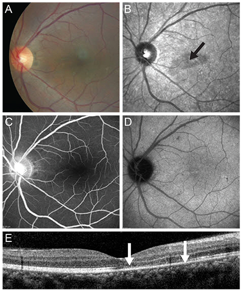
Fig. 1 Left eye of case 1 at presentation. (A) Color fundus photo showing retinal dot lesions around the macula. (B) Red-free fundus photograph revealing hypofluorescence in the macula (black arrow). (C) Fluorescence blockage by retinal hemorrhage can be seen in the fluorescein angiography. (D) Indocyanine green angiography shows retinal hemorrhage around the macula with hypofluorescence in the late phase. (E) Spectral domain optical coherence tomography images show foveal disruption of the photoreceptor layer (between the white arrows), whereas the external limiting membrane is continuous.
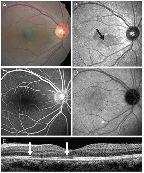
Fig. 2 Right eye of case 1 at presentation. (A) Color fundus photo showing retinal dot lesions around the macula. (B) Red-free fundus photograph revealing hypofluorescence in the macula (black arrow). (C) Fluorescence blockage by retinal hemorrhage can be seen in the fluorescein angiography. (D) Indocyanine green angiography shows retinal hemorrhage around the macula with hypofluorescence in the late phase. (E) Spectral domain optical coherence tomography images show foveal disruption of the photoreceptor layer (between the white arrows), whereas the external limiting membrane is continuous.
Reference
-
1. Gubler DJ. The global emergence/resurgence of arboviral diseases as public health problems. Arch Med Res. 2002; 33:330–342.2. Richardson S. Ocular symptoms and complications observed in dengue. Trans Am Ophthalmol Soc. 1933; 31:450–477.3. Cruz-Villegas V, Berrocal AM, Davis JL. Bilateral choroidal effusions associated with dengue fever. Retina. 2003; 23:576–578.4. Wen KH, Sheu MM, Chung CB, et al. The ocular fundus findings in dengue fever. Gaoxiong Yi Xue Ke Xue Za Zhi. 1989; 5:24–30.5. Chia A, Luu CD, Mathur R, et al. Electrophysiological findings in patients with dengue-related maculopathy. Arch Ophthalmol. 2006; 124:1421–1426.6. Morens DM. Antibody-dependent enhancement of infection and the pathogenesis of viral disease. Clin Infect Dis. 1994; 19:500–512.7. Su DH, Bacsal K, Chee SP, et al. Prevalence of dengue maculopathy in patients hospitalized for dengue fever. Ophthalmology. 2007; 114:1743–1747.8. Lim WK, Mathur R, Koh A, et al. Ocular manifestations of dengue fever. Ophthalmology. 2004; 111:2057–2064.9. Kurane I, Rothman AL, Livingston PG, et al. Immunopathologic mechanisms of dengue hemorrhagic fever and dengue shock syndrome. Arch Virol Suppl. 1994; 9:59–64.10. Lei HY, Yeh TM, Liu HS, et al. Immunopathogenesis of dengue virus infection. J Biomed Sci. 2001; 8:377–388.11. Lin CF, Lei HY, Shiau AL, et al. Antibodies from dengue patient sera cross-react with endothelial cells and induce damage. J Med Virol. 2003; 69:82–90.12. Iida T, Spaide RF, Kantor J. Retinal and choroidal arterial occlusion in Wegener's granulomatosis. Am J Ophthalmol. 2002; 133:151–152.
- Full Text Links
-
- Actions
-
Cited
- CITED
-
- Close
- Share
-
- Similar articles
-
- Optical Coherence Tomography to Evaluate Dengue
- Influence of RNFL Thickness on Visual Acuity and Visual Field in Bilateral Temporal Optic Atrophy
- Correlation of Preoperative Optical Coherence Tomography with Preand Postoperative Visual Acuity of Macular Hole
- The Usefulness of Optical Coherence Tomography in Macula-off Retinal Detachment
- Correlation between Foveal Thickness and Visual Acuity in Unilateral Resolved Central Serous Chorioretinopathy
