Histiocyte-Rich Reactive Lymphoid Hyperplasia of the Liver: Unusual Morphologic Features
- Affiliations
-
- 1Department of Pathology, Chonbuk National University, Medical School, Institute for Medical Sciences and the Center for Healthcare Technology Development, Jeonju, Korea. mws@chonbuk.ac.kr
- 2Department of Radiology, Chonbuk National University, Medical School, Institute for Medical Sciences and the Center for Healthcare Technology Development, Jeonju, Korea.
- 3Department of Surgery, Chonbuk National University, Medical School, Institute for Medical Sciences and the Center for Healthcare Technology Development, Jeonju, Korea.
- KMID: 1786866
- DOI: http://doi.org/10.3346/jkms.2008.23.1.156
Abstract
- Reactive lymphoid hyperplasia (RLH) of the liver is a rare entity and has also been termed nodular lymphoid lesion or pseudolymphoma of the liver. We report a case of hepatic RLH exhibiting unusual histiocyte-rich histologic features in a 47-yr-old woman in conjunction with a renal cell carcinoma. A follow-up computed tomography scan was done 14 months after a right radical nephrectomy for renal cell carcinoma revealed a nodular lesion in segment 5 of the liver. The lesion was interpreted as metastatic renal cell carcinoma or hepatocellular carcinoma based on the history of the patient and radiologic findings. Wedge resection of segment 5 was done with sufficient distance from the mass. Microscopically, the lesion was composed predominantly of peculiar histiocytic proliferation and was characterized by lymphoid aggregates forming a lymphoid follicle with germinal centers. The present case and prior cases reported in the literature suggest that RLH of the liver appear to be a heterogenous group of reactive inflammatory lesions that are often associated with autoimmune disease or malignant tumors.
MeSH Terms
Figure
-
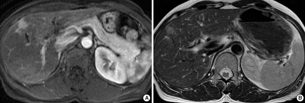
Fig. 1 (A) The respiratory-triggered T2-weighted turbo spin-echo MR imaging shows a small mass with intermediate high signal intensity (arrow) in liver segment 5. (B) The gadolinium-enhanced arterial phase MR imaging shows small bright nodular enhancement (arrow) in the same location as in A.
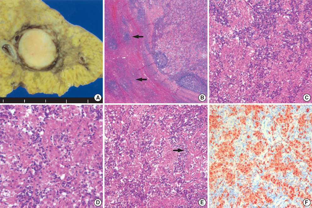
Fig. 2 (A) A cut section of the resected liver shows a well-circumscribed, yellowish-white mass in segment 5. (B) The lesion is composed predominantly of peculiar histiocytic proliferation and lymphoid aggregate forming lymphoid follicles on the edge of the nodule. Note the dense infiltrate of lymphocytes in several portal tracts immediately around the nodule (arrows, ×20, H&E). (C, D) The lesion is mainly composed of aggregates of epithelioid histiocytes simulating non-caseating granuloma (C ×200, D ×400, H&E). (E) Hyalinized structures in the nodule (arrow, ×200, H&E). (F) CD68 immunostaining highlights the large number of histiocytes (×200, CD68).
Reference
-
1. Isobe H, Sakamoto S, Sakai H, Masumoto A, Sonoda T, Adachi E, Nawata H. Reactive lymphoid hyperplasia of the liver. J Clin Gastroenterol. 1993. 16:240–244.
Article2. Sharifi S, Murphy M, Loda M, Pinkus GS, Khettry U. Nodular lymphoid lesion of the liver: an immune-mediated disorder mimicking lowgrade malignant lymphoma. Am J Surg Pathol. 1999. 23:302–308.3. Takahashi H, Sawai H, Matsuo Y, Funahashi H, Satoh M, Okada Y, Inagaki H, Takeyama H, Manabe T. Reactive lymphoid hyperplasia of the liver in a patient with colon cancer: report of two cases. BMC Gastroenterol. 2006. 6:–25.
Article4. Willenbrock K, Kriener S, Oeschger S, Hansmann ML. Nodular lymphoid lesion of the liver with simultaneous focal nodular hyperplasia and hemangioma: discrimination from primary hepatic MALT-type non-Hodgkin's lymphoma. Virchows Arch. 2006. 448:223–227.
Article5. Grouls V. Pseudolymphoma (inflammatory pseudotumor) of the liver. Zentralbl Allg Pathol. 1987. 133:565–568.6. Ohtsu T, Sasaki Y, Tanizaki H, Kawano N, Ryu M, Satake M, Hasebe T, Mukai K, Fujikura M, Tamai M, Abe K. Development of pseudolymphoma of liver following interferon-alpha therapy for chronic hepatitis B. Intern Med. 1994. 33:18–22.
Article7. Katayanagi K, Terada T, Nakanuma Y, Ueno T. A case of pseudolymphoma of the liver. Pathol Int. 1994. 44:704–711.
Article8. Kim SR, Hayashi Y, Kang KB, Soe CG, Kim JH, Yang MK, Itoh H. A case of pseudolymphoma of the liver with chronic hepatitis C. J Hepatol. 1997. 26:209–214.
Article9. Okubo H, Maekawa H, Ogawa K, Wada R, Sekigawa I, Iida N, Maekawa T, Hashimoto H, Sato N. Pseudolymphoma of the liver associated with Sjogren's syndrome. Scand J Rheumatol. 2001. 30:117–119.10. Nagano K, Fukuda Y, Nakano I, Katano Y, Toyoda H, Nonami T, Nagasaka T, Hayakawa T. Reactive lymphoid hyperplasia of liver coexisting with chronic thyroiditis: radiographical characteristics of the disorder. J Gastroenterol Hepatol. 1999. 14:163–167.11. Pantanowitz L, Saldinger PF, Kadin ME. Pathologic quiz case: Hepatic mass in a patient with renal cell carcinoma. Arch Pathol Lab Med. 2001. 125:577–578.
Article12. Maehara N, Chijiiwa K, Makino I, Ohuchida J, Kai M, Kondo K, Moriguchi S, Marutsuka K, Asada Y. Segmentectomy for reactive lymphoid hyperplasia of the liver: report of a case. Surg Today. 2006. 36:1019–1023.
Article13. Sato K, Ueda Y, Yokoi M, Hayashi K, Kosaka T, Katsuda S. Reactive lymphoid hyperplasia of the liver in a patient with multiple carcinomas: a case report and brief review. J Clin Pathol. 2006. 59:990–992.
Article14. Tanizawa T, Eishi Y, Kamiyama R, Nakahara M, Abo Y, Sumita T, Kawano N. Reactive lymphoid hyperplasia of the liver characterized by an angiofollicular pattern mimicking Castleman's disease. Pathol Int. 1996. 46:782–786.
Article
- Full Text Links
-
- Actions
-
Cited
- CITED
-
- Close
- Share
-
- Similar articles
-
- Primary Cutaneous T-cell/histiocyte-rich B-cell Lymphoma
- Reactive Lymphoid Hyperplasia Treated with Radiofrequency Ablation
- Orbital Lymphocytic Tumor Gradually Progressed to More Malignant Form during 4 Years
- Diagnostic Pediatric Colonoscopy for Lymphoid Hyperplasia of Terminal Ileum
- Reactive lymphoid hyperplasia of the liver
