Morphological variations and accessory ossicles in the peroneal and tibialis muscles
- Affiliations
-
- 1Division of Anatomy, The Ohio State University College of Medicine, Columbus, OH, USA.
- 2School of Health and Rehabilitations Sciences, The Ohio State University College of Medicine, Columbus, OH, USA. Laura.Boucher@osumc.edu
- KMID: 2459552
- DOI: http://doi.org/10.5115/acb.19.030
Abstract
- This study describes five bilateral anatomical variations in the feet of a 97-year-old male cadaver. Following routine dissection, all variants were measured and documented. Three accessory tendons and two accessory ossicles were identified. Bilateral accessory tendons were present from the tibialis anterior (type II), peroneus tertius (type III), and peroneus brevis muscles. Accessory tendon length was 36-104 mm and width was 1-3 mm each inserting more distally then the main tendon. Accessory ossicles were identified as an accessory navicular and os peroneum, respectively. Individually, each variation has varying prevalence rates in the literature, but to date, no known studies have been published describing the combined presence of all five bilateral variations. The acknowledgement of multi-variant cases such as this one may be helpful in the clinical setting, particularly for patients with pathology or for those undergoing foot and ankle surgery.
Figure
-
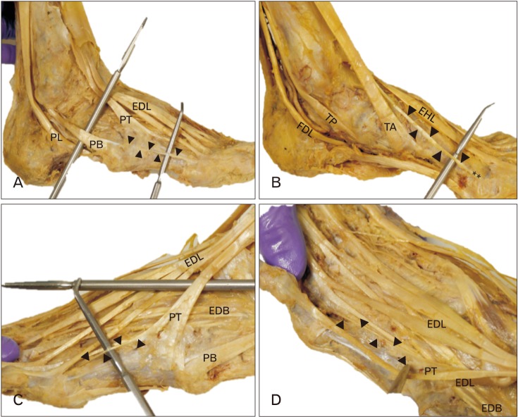
Fig. 1 (A) Accessory tendon morphology. The accessory tendon of peroneus brevis muscle (PB) (arrowheads) extended from the main tendon of PB to attach laterally on the head of the fifth metatarsal. Also visible are the tendons of peroneus longus (PL), peroneus tertius (PT), and extensor digitorum longus (EDL). (B) The main tendon of the tibialis anterior (TA) inserts on the medial cuneiform and first metatarsal base, while the accessory tendon of TA (arrowheads) inserts at the head of the first metatarsal (**). Also visible is the tendon of extensor hallucis longus (EHL), the tendon of tibialis posterior muscle (TP), and the tendon of flexor digitorum longus (FDL). (C, D) Anomalous bifurcation in the tendon of PT indicated by black arrowheads. Image C demonstrates the lateral view of the tendon, while Image D demonstrates the dorsolateral view of the tendon as it attached to the lateral base of the fourth proximal phalanx. Also visible are the muscle belly of extensor digitorum brevis (EDB), tendon of PB, and tendons of EDL.

Fig. 2 (A) Ossicle morphology. The os peroneum (OP, *) within the tendon of peroneus longus (PL), adjacent to the cuboid. Arrow indicates the convex facet on the cuboid bone at the site of articulation with the sesamoid bone. Also visible is the tendon of peroneus brevis muscle (PB). (B) Isolated OP, measuring 20 mm long and 13 mm wide. (C) Accessory navicular (AN; indicated by tip of probe) located within the tendon of tibialis posterior muscle (TP), proximal to its division and adjacent to the navicular tuberosity (NT). Also visible are the long plantar ligament (LPL), short plantar ligament (SPL), and spring ligament (SL). (D) Isolated AN, measuring 13 mm long and 7 mm wide.
Cited by 2 articles
-
Dimensions of pes anserinus of the lower extremity, an anatomical study with its surgical implications
Rajanigandha Vadgaonkar, M.D. Prameela, Chettiar Ganesh Kumar, Vandana Blossom, Mamatha Tonse, B.V. Murlimanju, Mangala M. Pai, Latha V. Prabhu
Anat Cell Biol. 2021;54(2):178-183. doi: 10.5115/acb.20.275.Concomitant variations of the tibialis anterior, and extensor hallucis longus, and extensor hallucis brevis muscles
Jenilkumar Patel, Graham Dupont, Joho Katsuta, Joe Iwanaga, Łukasz Olewnik, R. Shane Tubbs
Anat Cell Biol. 2023;56(1):137-140. doi: 10.5115/acb.22.009.
Reference
-
1. Ercikti N, Apaydin N, Kocabiyik N, Yazar F. Insertional characteristics of the peroneus tertius tendon: revisiting the anatomy of an underestimated m uscle. J Foot Ankle Surg. 2016; 55:709–713. PMID: 26860045.2. Luchansky E, Paz Z. Variations in the insertion of tibialis anterior muscle. Anat Anz. 1986; 162:129–136. PMID: 3789411.3. Imre N, Kocabiyik N, Sanal HT, Uysal M, Ozan H, Yazar F. The peroneus brevis tendon at its insertion site on fifth metatarsal bone. Foot Ankle Surg. 2016; 22:41–45. PMID: 26869499.4. Nwawka OK, Hayashi D, Diaz LE, Goud AR, Arndt WF 3rd, Roemer FW, Malguria N, Guermazi A. Sesamoids and accessory ossicles of the foot: anatomical variability and related pathology. Insights Imaging. 2013; 4:581–593. PMID: 24006205.5. Joshi SD, Joshi SS, Athavale SA. Morphology of peroneus tertius muscle. Clin Anat. 2006; 19:611–614. PMID: 16317742.6. Demir BT, Gümüşalan Y, Üzel M, Çevik HB. The variations of peroneus digiti quinti muscle and its contribution to the extension of the fifth toe. A cadaveric study. Saudi Med J. 2015; 36:1285–1289. PMID: 26593160.7. Yammine K. The accessory peroneal (fibular) muscles: peroneus quartus and peroneus digiti quinti. A systematic review and meta-analysis. Surg Radiol Anat. 2015; 37:617–627. PMID: 25638531.8. Musial WW. Variations of the terminal insertions of the anterior and posterior tibial muscles in man. Folia Morphol. 1963; 26:237–247.9. Benninger B, Kloenne J. The clinical importance of the os peroneum: a dissection of 156 limbs comparing the incidence rates in cadavers versus chronological roentgenograms. Foot Ankle Online J. 2011; 2:2.10. Bareither DJ, Muehleman CM, Feldman NJ. Os tibiale externum or sesamoid in the tendon of tibialis posterior. J Foot Ankle Surg. 1995; 34:429–434. PMID: 8590876.11. Sonmez M, Kosar I, Cimen M. The supernumerary peroneal muscles: case report and review of the literature. Foot Ankle Surg. 2000; 6:125–129.12. Raheja S, Choudhry R, Singh P, Tuli A, Kumar H. Morphological description of combined variation of distal attachments of fibulares in a foot. Surg Radiol Anat. 2005; 27:158–160. PMID: 15580345.13. Fidziańska A, Goebel HH. Human ontogenesis. 3. Cell death in fetal muscle. Acta Neuropathol. 1991; 81:572–577. PMID: 1858485.14. Webb JN. The development of human skeletal muscle with particular reference to muscle cell death. J Pathol. 1972; 106:221–228. PMID: 5046933.15. Webb JN. Cell death in developing skeletal muscle: histochemistry and ultrastructure. J Pathol. 1977; 123:175–180. PMID: 592022.
- Full Text Links
-
- Actions
-
Cited
- CITED
-
- Close
- Share
-
- Similar articles
-
- Clinical Features and Radiological Differential Diagnoses of Symptomatic Sesamoid Bones and Accessory Ossicles: A Pictorial Essay
- Prevalence of Accessory Bones and Tarsal Coalitions Based on Radiographic Findings in a Healthy, Asymptomatic Population
- The accessory deep peroneal nerve : frequency and electrophysiological findings
- A Case of Traumatic Dislocation of PeroneaI Tendons
- A case of neonatal peroneal neuropathy with intrauterine onset
