Primary Synovial Sarcoma of the Thyroid Gland
- Affiliations
-
- 1Department of Pathology, College of Medicine, Hanyang University, Seoul, Korea. parkmh@hanyang.ac.kr
- 2Department of Otolaryngology-Head and Neck Surgery, College of Medicine, Hanyang University, Seoul, Korea.
- 3Department of Pathology, Asan Medical Center, College of Medicine, Ulsan University, Seoul, Korea.
- KMID: 1785806
- DOI: http://doi.org/10.3346/jkms.2007.22.S.S154
Abstract
- Synovial sarcoma is a rare but distinct soft tissue neoplasm, most commonly occurring in para-articular regions of the extremities of young adults and also occurring in the head and neck region. To the best of our knowledge, only one case of primary synovial sarcoma of the thyroid has been previously reported. Here, we report a 15-yr-old man who had a chief complaint of a palpable neck mass. The neck computed tomography revealed a relatively well-demarcated solid mass in the left thyroid gland. After fine needle aspiration cytology, total thyroidectomy and lymph node dissection were performed. Grossly, the mass was covered by the same capsule as the thyroid gland, measuring 6X5X5 cm in dimensions and weighing 78 gm. The cut surface showed a well demarcated, lobulated, grayish tan, and rubbery solid tumor. Histologically, this tumor was a biphasic synovial sarcoma. Immunohistochemical, ultrastructural, genetic studies, and cytologic findings were all consistent with synovial sarcoma. When synovial sarcomas arise in this unusual site, recognition and differential diagnosis become more difficult. The differential diagnosis of a spindle epithelial tumor with thymus-like differentiation is very difficult due to their similar clinical, histological, and immunohistochemical features. Ultrastructural and cytogenetic studies for synovial sarcoma are necessary to establish a definitive diagnosis.
Keyword
MeSH Terms
Figure
-

Fig. 1 The neck computed tomography reveals a well-demarcated solid mass at the superior and lateral aspect of the left thyroid gland, suggesting either a primary thyroid mass or soft tissue tumor.
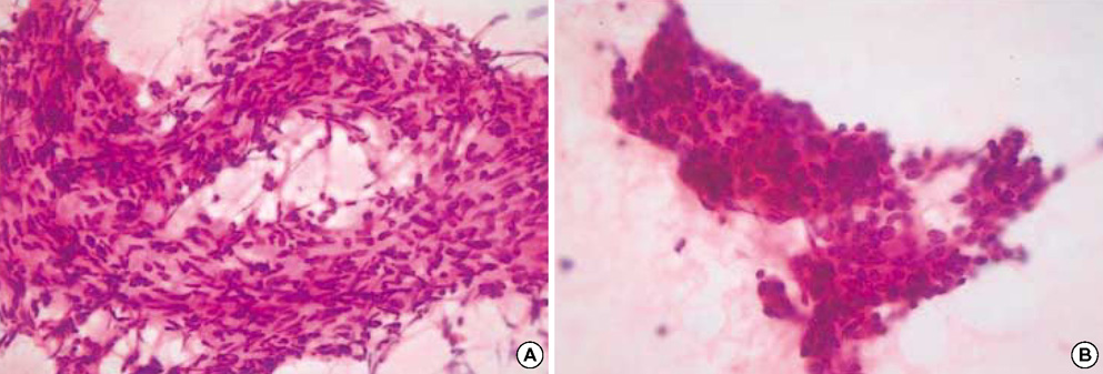
Fig. 2 The fine needle aspiration cytology shows several cell clusters, which are composed of spindle-shaped (A) or epithelioid cells (B). The nuclei are round to oval and spindle-shaped and show finely granular chromatin with a moderate amount of pale eosinophilic cytoplasm.

Fig. 3 A capsule covers the mass and the thyroid gland, and the cut surface of the mass shows a well-demarcated, lobulated and tan-colored solid mass with rubbery consistency.
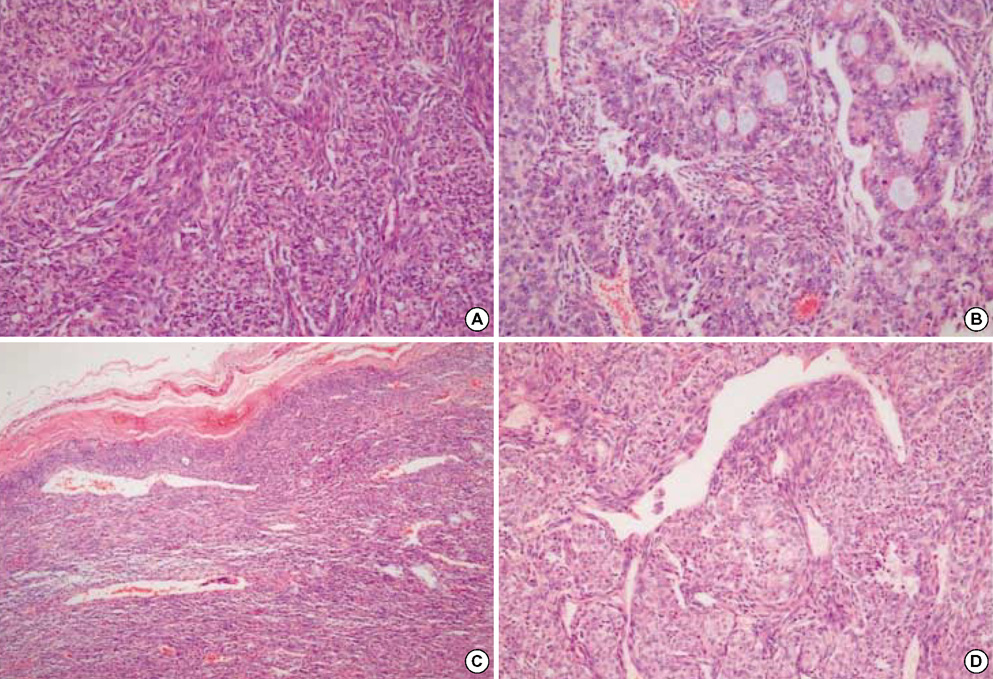
Fig. 4 Microscopically, the tumor shows a biphasic growth pattern (A). The spindle cell component consists of fascicles of atypical fibroblast-like cells, and the epithelial component is composed of solid nests of epithelioid cells with well-formed glandular structures (B). The periphery of the tumor shows more densely packed spindle cells with calcification (C). A focal hemangiopericytic growth pattern is present (D).
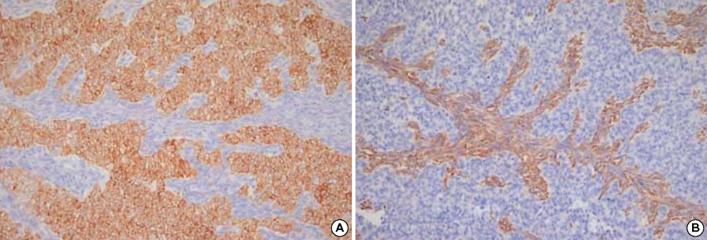
Fig. 5 On immunohistochemistry, the epithelial cells are diffusely and strongly stained with cytokeratin (A), whereas the spindle cells are diffusely stained with vimentin (B).

Fig. 6 On ultrastructural examination, the tumor is composed of solid nests of epithelial cells and fascicles of spindle cells, which are separated by basal lamina (A, arrows). The epithelial cells have desmosome-like cell junctions (B, arrowhead).
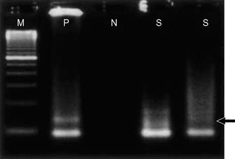
Fig. 7 The SYT-SSX fusion gene transcript was detected (arrow) in paraffin-embedded tumor tissue using the reverse transcriptase-polymerase chain reaction method. (Lane M, 100-bp ladder DNA marker; lane P, positive control; lane N, negative control; lane S, tumor tissue)
Cited by 1 articles
-
Synovial Sarcoma of the Thyroid Gland
Chang Hwan Ryu, Kyung-Ja Cho, Seung-Ho Choi
Clin Exp Otorhinolaryngol. 2011;4(4):204-206. doi: 10.3342/ceo.2011.4.4.204.
Reference
-
1. Weiss SW, Goldblum JR, Enzinger FM. Enzinger and Weiss's Soft Tissue Tumors. 2001. St. Louis, USA: Mosby;1483–1509.2. Raney RB. Synovial sarcoma in young people: background, prognostic factors, and therapeutic questions. J Pediatr Hematol Oncol. 2005. 27:207–211.3. Nielsen GP, Shaw PA, Rosenberg AE, Dickersin GR, Young RH, Scully RE. Synovial sarcoma of the vulva: a report of two cases. Mod Pathol. 1996. 9:970–974.4. Smith CJ, Ferrier AJ, Russell P, Danieletto S. Primary synovial sarcoma of the ovary: first reported case. Pathology. 2005. 37:385–387.
Article5. Holla P, Hafez GR, Slukvin I, Kalayoglu M. Synovial sarcoma, a primary liver tumor--a case report. Pathol Res Pract. 2006. 202:385–387.6. Suster S, Moran CA. Primary synovial sarcomas of the mediastinum: a clinicopathologic, immunohistochemical, and ultrastructural study of 15 cases. Am J Surg Pathol. 2005. 29:569–578.7. Kikuchi I, Anbo J, Nakamura S, Sugai T, Sasou S, Yamamoto M, Oda Y, Shiratsuchi H, Tsuneyoshi M. Synovial sarcoma of the thyroid. Report of a case with aspiration cytology findings and gene analysis. Acta Cytol. 2003. 47:495–500.8. Miettinen M, Virtanen I. Synovial sarcoma--a misnomer. Am J Pathol. 1984. 117:18–25.9. Dickersin GR. Synovial sarcoma: a review and update, with emphasis on the ultrastructural characterization of the nonglandular component. Ultrastruct Pathol. 1991. 15:379–402.
Article10. Pilch BZ. Head and Neck Surgical Pathology. 2001. Philadelphia, USA: Lippincott Williams & Wilkins;365–371.11. Kirby PA, Ellison WA, Thomas PA. Spindle epithelial tumor with thymus-like differentiation (SETTLE) of the thyroid with prominent mitotic activity and focal necrosis. Am J Surg Pathol. 1999. 23:712–716.
Article12. Tsuji S, Hisaoka M, Morimitsu Y, Hashimoto H, Shimajiri S, Komiya S, Ushijima M, Nakamura T. Detection of SYT-SSX fusion transcripts in synovial sarcoma by reverse transcription-polymerase chain reaction using archival paraffin-embedded tissues. Am J Pathol. 1998. 153:1807–1812.
Article
