Anaplastic Ganglioglioma in a Middle-aged Woman: A Case Report with a Review of the Literature
- Affiliations
-
- 1Department of Neurosurgery, Institute of Health Science, Gyeongsang National University School of Medicine, Jinju, Korea. gnuhjjm@nongae.gsnu.ac.kr
- KMID: 1785803
- DOI: http://doi.org/10.3346/jkms.2007.22.S.S139
Abstract
- We report a case of anaplastic ganglioglioma. A 45-yr-old woman was admitted with a 5-month history of headache and dizziness, both of which progressed slowly. Preoperative magnetic resonance imaging revealed a strong enhancing mass in the left frontal lobe extending to the cingulate gyrus. Adjuvant radiation therapy and chemotherapy were given after gross total resection of the tumor. Histological and immunohistochemical studies showed an anaplastic ganglioglioma. Gangliogliomas of the central nervous system are rather uncommon tumors, and anaplastic ones are extremely rare. The pertinent literature regarding gangliogliomas is reviewed.
Keyword
MeSH Terms
Figure
-
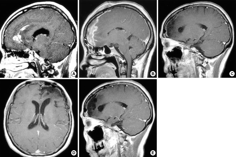
Fig. 1 Preoperative MRI showing an irregular strong enhancing mass in the left frontal lobe extending to the cingulate gyrus (A). MRI on the 12th postoperative day showing multiple irregularly shaped enhancing lesions along the resection margin of the left frontal lobe and cingulate gyrus (B). After chemotherapy and radiation therapy, MRI 5 months postoperatively revealed that the irregular enhancing lesions in the resected margin of the left frontal lobe and cingulate gyrus had disappeared (C). And, the last follow-up MRI at 35 months postoperatively shows no evidence of recurrence (D, E).
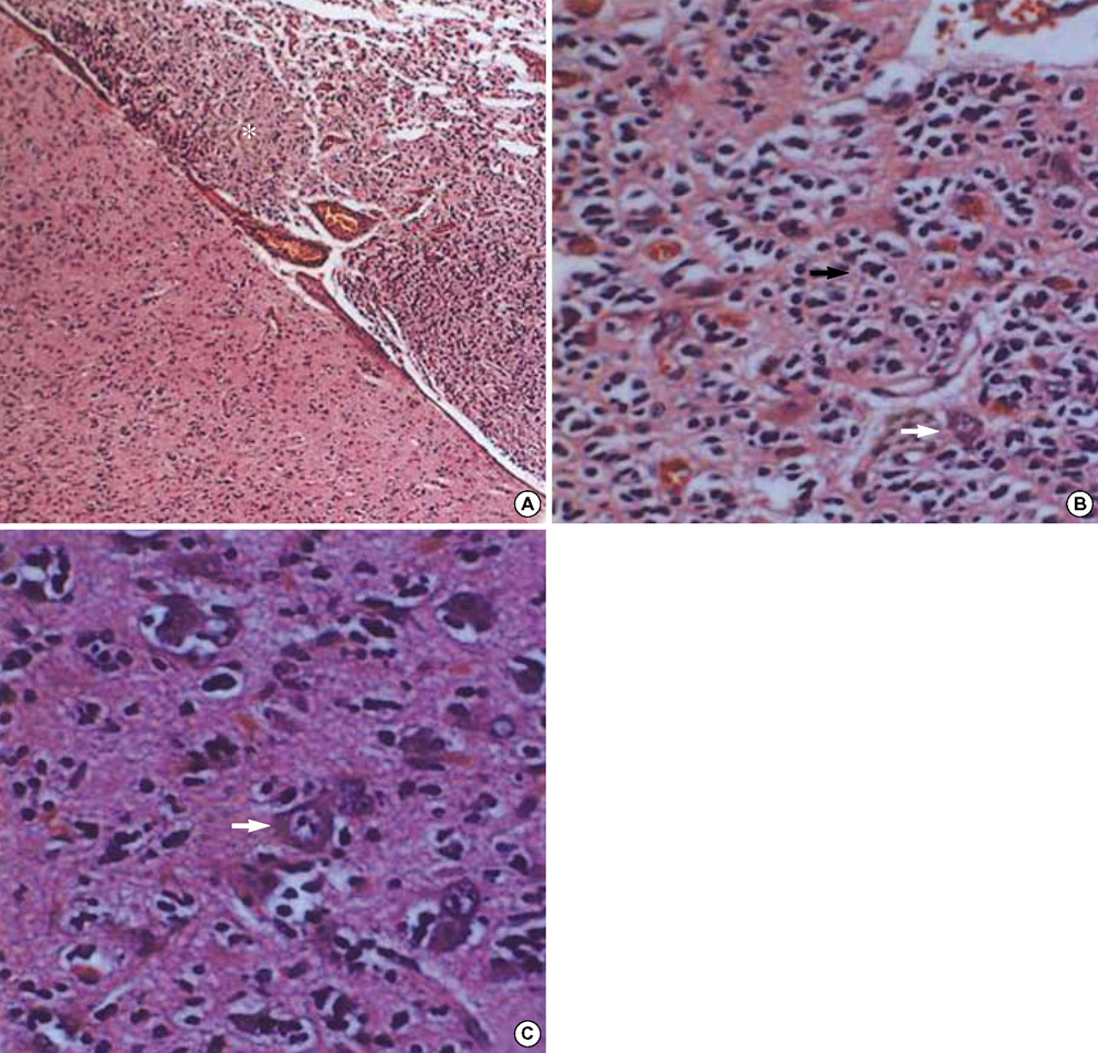
Fig. 2 Photomicrographs showing the histopathology of the ganglioglioma in our case. (A) The tumor cells infiltrated the gray matter and subarachnoid space. The tumor cells in the gray matter consisted of neoplastic ganglion cells and glial cells. Those in the subarachnoid space (asterisk) are mainly glial cells and show increased cellularity, nuclear atypism, and occasional mitosis (hematoxylin and eosin, original magnification, ×40). (B) Atypical glial cells (arrow) and scattered ganglion cells (white arrow) constitute the ganglioglioma. Note the increased cellularity and nuclear atypism of the glial cells (hematoxylin and eosin, original magnification, ×400). (C) Neoplastic ganglion cells (arrow) are clustered abnormally and lack orientation and polarity. Most have large nuclei with prominent nucleoli (hematoxylin and eosin, original magnification, ×400).

Fig. 3 Immunostaining features suggestive of anaplastic gangliogliomas. Neoplastic ganglion cells are positive for NSE (A) and synaptophysin (B). (C) Glial components are positive for GFAP, and scattered ganglion cells are negative. (D) The Ki-67 LI is high (20%) in the region of anaplastic transformation.
Reference
-
1. Russell DS, Rubinstein LJ. Pathology of tumors of the nervous system. 1989. 5th Ed. Baltimore, USA: Williams & Wilkins;289–307.2. Haddad SF, Moore SA, Menezes AH, VanGilder JC. Ganglioglioma: 13 years of experience. Neurosurgery. 1992. 31:171–178.3. Hall WA, Yunis EJ, Albright AL. Anaplastic ganglioglioma in an infant: case report and review of the literature. Neurosurgery. 1986. 19:1016–1020.
Article4. Kitano M, Takayama S, Nagao T, Yoshimura O. Malignant ganglioglioma of the spinal cord. Acta Pathol Jpn. 1987. 37:1009–1018.
Article5. Lang FF, Epstein FJ, Ransohoff J, Allen JC, Wisoff J, Abbott IR, Miller DC. Central nervous system gangliogliomas. Part 2: clinical outcome. J Neurosurg. 1993. 79:867–873.6. Wolf HK, Muller MB, Spanle M, Zentner J, Schramm J, Wiestler OD. Ganglioglioma: a detailed histopathological and immunohistochemical analysis of 61 cases. Acta Neuropathol (Berl). 1994. 88:166–173.
Article7. Johannsson JH, Rekate HL, Roessmann U. Gangliogliomas: pathological and clinical correlation. J Neurosurg. 1981. 54:58–63.
Article8. Jay V, Squire J, Becker LE, Humphreys R. Malignant transformation in a ganglioglioma with anaplastic neuronal and astrocytic components: report of a case with flow cytometric and cytogenetic analysis. Cancer. 1994. 73:2862–2868.
Article9. Matsuzaki K, Uno M, Kageji T, Hirose T, Nagahiro S. Anaplastic ganglioglioma of the cerebellopontine angle. Case report. Neurol Med Chir (Tokyo). 2005. 45:591–595.10. Di Patre PL, Payer M, Brunea M, Delavelle J, De Tribolet N, Pizzolato G. Malignant transformation of a spinal cord ganglioglioma--case report and review of the literature. Clin Neuropathol. 2004. 23:298–303.11. Mittler MA, Walters BC, Fried AH, Sotomayor EA, Stopa EG. Malignant glial tumor arising from the site of a previous hamartoma/ganglioglioma: coincidence or malignant transformation? Pediatr Neurosurg. 1999. 30:132–134.
Article12. Hirose T, Kannuki S, Nishida K, Matsumoto K, Sano T, Hizawa K. Anaplastic ganglioglioma of the brain stem demonstrating active neurosecretory features of neoplastic neuronal cells. Acta Neuropathol (Berl). 1992. 83:365–370.
Article13. Blümcke I, Wiestler OD. Gangliogliomas: an intriguing tumor entity associated with focal epilepsies. J Neuropathol Exp Neurol. 2002. 61:575–584.
Article14. Allegranza A, Pileri S, Frank G, Ferracini R. Cerebral ganglioglioma with anaplastic oligodendroglial component. Histopathology. 1990. 17:439–441.
Article15. Araki M, Fan J, Haraoka S, Moritake T, Yoshii Y, Watanabe T. Extracranial metastasis of anaplastic ganglioglioma through a ventriculoperitoneal shunt: a case report. Pathol Int. 1999. 49:258–263.
Article16. Campos MG, Zentner J, Ostertun B, Wolf HK, Schramm J. Anaplastic ganglioglioma: case report and review of the literature. Neurol Res. 1994. 16:317–320.
Article17. Danjoux M, Sabatier J, Uro-Coste E, Roche H, Delisle MB. Anaplastic temporal ganglioglioma with spinal metastasis. Ann Pathol. 2001. 21:55–58.18. Nair V, Suri VS, Tatke M, Saran RK, Malhotra V, Singh D. Gangliogliomas: a report of five cases. Indian J Cancer. 2004. 41:41–46.19. Suzuki H, Otsuki T, Iwasaki Y, Katakura R, Asano H, Tadokoro M, Suzuki Y, Tezuka F, Takei H. Anaplastic ganglioglioma with sarcomatous component: an immunohistochemical study and molecular analysis of p53 tumor suppressor gene. Neuropathology. 2002. 22:40–47.20. Kurian NI, Nair S, Radhakrishnan VV. Anaplastic ganglioglioma: case report and review of literature. Br J Neurosurg. 1998. 12:277–280.21. Sasaki A, Hirato J, Nakazato Y, Tamura M, Kadowaki H. Recurrent anaplastic ganglioglioma: pathological characterization of tumor cells. Case report. J Neurosurg. 1996. 84:1055–1059.22. Whittle IR, Mitchener A, Atkinson HD, Wharton SB. Anaplastic progression in low grade glioneural neoplasms. Acta Neuropathol (Berl). 2002. 104:215.
Article23. Demierre B, Stichnoth FA, Hori A, Spoerri O. Intracerebral ganglioglioma. J Neurosurg. 1986. 65:177–182.
Article24. Park SH, Vinters HV. Ganglion cell tumors. Korean J Pathol. 2002. 36:167–174.
- Full Text Links
-
- Actions
-
Cited
- CITED
-
- Close
- Share
-
- Similar articles
-
- Rapidly Growing Anaplastic Ganglioglioma Mimicking Brain Metastasis in a Middle-Aged Woman: A Case Report
- Undiagnosed Anaplastic Ganglioglioma Resulting in the Sudden Unexpected Death of a Young Woman
- Ganglioglioma: A Case Report
- Desmoplastic Infantile Ganglioglioma: A Case Report
- Cervical Ganglioglioma
