J Korean Med Sci.
2007 Sep;22(Suppl):S129-S133. 10.3346/jkms.2007.22.S.S129.
A Magnetic Resonance-based Seed Localization Method for I-125 Prostate Implants
- Affiliations
-
- 1Department of Radiation Oncology, Ewha Womans University, Seoul, Korea. renalee@ewha.ac.kr
- 2Department of Radiology, Ewha Womans University, Seoul, Korea.
- 3Department of Radiation Oncology, Gachon Medical School, Gil Medical Center, Incheon, Korea.
- 4Department of Radiation Oncology, Yeungnam University, Daegu, Korea.
- KMID: 1785801
- DOI: http://doi.org/10.3346/jkms.2007.22.S.S129
Abstract
- This study was performed to develop and evaluate a semi-automatic seed localization algorithm from magnetic resonance (MR) images for interstitial prostate brachytherapy. The computerized tomography (CT) and MR images (3 mm-slice thickness) of six patients who had received real-time MR imaging-guided interstitial prostate brachytherapy were obtained. An automatic seed localization method was performed on CT images to obtain seed coordinates, and an algorithm for seed localization from MR images of the prostate was developed and tested. The resultant seed distributions from MR images were then compared to CT-derived distribution by matching the same seeds and calculating percent volume receiving 100% of the prescribed dose and the extent of the volume in 3-dimensions. The semiautomatic seed localization method made it possible to extract more than 90% of the seeds with either less than 8% of noises or 3% of missing seeds. The mean volume difference obtained from CT and MR receiving 100% of the prescribed dose was less than 3%. The maximum extent of the volume receiving the prescribed dose were 0.3, 0.6, and 0.2 cm in x, y, and z directions, respectively. These results indicate that the algorithm is very useful in identifying seeds from MR image for post-implant dosimety.
Keyword
MeSH Terms
-
Algorithms
Brachytherapy/*methods/statistics & numerical data
Humans
Iodine Radioisotopes/*administration & dosage
*Magnetic Resonance Imaging, Interventional/statistics & numerical data
Male
Prostatic Neoplasms/*pathology/radiography/*radiotherapy
Radiotherapy Dosage
Radiotherapy Planning, Computer-Assisted
Tomography, X-Ray Computed/statistics & numerical data
Tumor Burden
Figure
-
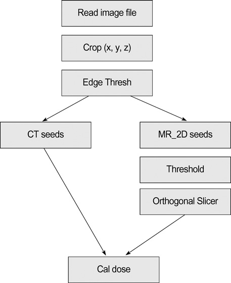
Fig. 1 Schematic diagram of seed localization algorithm.
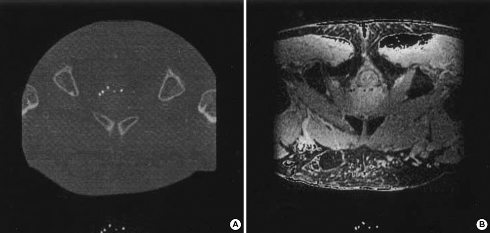
Fig. 2 Representative (A) computed tomogram (CT) and (B) magnetic resonance (MR) images. Seeds are depicted as white dots on CT and signal void on the T2-weighted MR image. Top images are original scans and bottom images are after seed extraction.
Reference
-
1. American Cancer Society. Cancer Facts and Figures. 1977. Atlanta, GA:2. Jemal A, Siegel R, Ward E, Murray T, Xu J, Smigal C, Thun MJ. Cancer statistics 2006. CA Cancer J Clin. 2006. 56:106–130.
Article3. Cho JH. Radiation Treatment for Prostate Cancer. J Korean Med Assoc. 2004. 47:424–431.
Article4. Stone NN, Stock RG. Long-term urinary, sexual, and rectal morbidity in patients treated with iodine-125 prostate brachytherapy followed up for a minimum of 5 years. Urology. 2007. 69:338–342.
Article5. Oh MM, Bahk YW, Tropper SE. Preliminary Report of Clinical Experience of Iodine-125 Seed Implant for Early Prostatic Cancer: The First Case in Korea. Korean J Urol. 2001. 42:1235–1240.6. Buron C, Le Vu B, Cosset JM, Pommier P, Peiffert D, Delannes M, Flam T, Guerif S, Salem N, Chauveinc L, Livartowski A. Brachytherapy versus prostatectomy in localized prostate cancer: results of a French multicenter prospective medico-economic study. Int J Radiat Oncol Biol Phys. 2007. 67:812–822.
Article7. Holm HH, Juul N, Pedersen JF, Hansen H, Strøyer I. Transperineal 125iodine seed implantation in prostatic cancer guided by transrectal ultrasonography. J Urol. 1983. 130:283–286.8. Xue J, Waterman F, Handler J, Gressen E. Localization of linked 125I seeds in postimplant TRUS images for prostate brachytherapy dosimetry. Int J Radiat Oncol Biol Phys. 2005. 62:912–919.
Article9. Horwitz EM, Mitra RK, Uzzo RG, Das IJ, Pinover WH, Hanlon AL, McNeeley SW, Hanks GE. Impact of target volume coverage with Radiation Therapy Oncology Group (RTOG) 98-05 guidelines for transrectal ultrasound guided permanent Iodine-125 prostate implants. Radiother Oncol. 2003. 66:173–179.
Article10. Koutrouvelis PG. Three-dimensional stereotactic posterior ischiorectal space computerized tomography guided brachytherapy of prostate cancer: a preliminary report. J Urol. 1998. 159:142–145.11. Tanaka O, Hayashi S, Kanematsu M, Matsuo M, Nakano M, Maeda S, Deguchi T, Hoshi H. CT-based postimplant dosimetry of prostate brachytherapy: comparison of 1-mm and 5-mm section CT. Radiat Med. 2007. 25:22–26.
Article12. Moerland MA, Wijrdeman HK, Beersma R, Bakker CJ, Battermann JJ. Evaluation of permanent I-125 prostate implants using radiography and magnetic resonance imaging. Int J Radiat Oncol Biol Phys. 1997. 37:927–933.
Article13. Dubois DF, Prestidge BR, Hotchkiss LA, Bice WS Jr, Prete JJ. Source localization following permanent transperineal prostate interstitial brachytherapy using magnetic resonance imaging. Int J Radiat Oncol Biol Phys. 1997. 39:1037–1041.
Article14. Amols HI, Rosen II. A three-film technique for reconstruction of radioactive seed implants. Med Phys. 1981. 8:210–214.
Article15. Haworth A, Ebert M, St Clair S, Carey BM, Flynn A, Bottomley DM, Duchesne GM, Joseph D, Ash D. Impact of selection of post-implant technique on dosimetry parameters for permanent prostate implants. Brachytherapy. 2005. 4:146–153.
Article16. Crook J, McLean M, Yeung I, Williams T, Lockwood G. MRI-CT fusion to assess postbrachytherapy prostate volume and the effects of prolonged edema on dosimetry following transperineal interstitial permanent prostate brachytherapy. Brachytherapy. 2004. 3:55–60.
Article17. Roy JN, Wallner KE, Harrington PJ, Ling CC, Anderson LL. A CT-based evaluation method for permanent implants: application to prostate. Int J Radiat Oncol Biol Phys. 1993. 26:163–169.
Article18. Brinkmann DH, Kline RW. Automated seed localization from CT datasets of the prostate. Med Phys. 1998. 25:1667–1672.
Article19. Roach M 3rd, Faillace-Akazawa P, Malfatti C, Holland J, Hricak H. Prostate volumes defined by magnetic resonance imaging and computerized tomographic scans for three-dimensional conformal radiotherapy. Int J Radiat Oncol Biol Phys. 1996. 35:1011–1018.
Article20. Smith WL, Lewis C, Bauman G, Rodrigues G, D'Souza D, Ash R, Ho D, Venkatesan V, Downey D, Fenster A. Prostate volume contouring: a 3D analysis of segmentation using 3DTRUS, CT, and MR. Int J Radiat Oncol Biol Phys. 2007. 67:1238–1247.
Article
- Full Text Links
-
- Actions
-
Cited
- CITED
-
- Close
- Share
-
- Similar articles
-
- A Study on Image Reconstruction for Seed Localization for Permanent Prostate Brachytherapy
- Medical imaging of prostate cancer
- Seed-Based Resting-State Functional MRI for Presurgical Localization of the Motor Cortex: A Task-Based Functional MRI-Determined Seed Versus an Anatomy-Determined Seed
- Preliminary Report of Clinical Experience of Iodine-125 Seed Implant for Early Prostatic Cancer: The First Case in Korea
- MR-Guided Targeted Prostate Biopsy from Radiologists’ Perspective
