J Korean Med Sci.
2004 Feb;19(1):62-68. 10.3346/jkms.2004.19.1.62.
Relationships between High-Resolution Computed Tomography, Lung Function and Bacteriology in Stable Bronchiectasis
- Affiliations
-
- 1Department of Internal Medicine, College of Medicine, Ewha Womans University, Seoul, Korea. hs1017@ewha.ac.kr
- 2Department of Diagnostic Radiology, Medical Research Center, College of Medicine, Ewha Womans University, Seoul, Korea.
- 3Department of Radiology., Kangbuk Samsung Hospital, Sungkyunkwan University School of Medicine, Samsung Medical Center, Seoul, Korea.
- KMID: 1785695
- DOI: http://doi.org/10.3346/jkms.2004.19.1.62
Abstract
- To determine the relationship between high-resolution computed tomography (HRCT) findings, lung function, and bacteriology in bronchiectasis, we conducted a retrospective study of 49 Korean patients with stable bronchiectasis. To quantify the extent and severity of bronchiectasis, we used a CT scoring system consisting of bronchial dilatation, bronchial wall thickening, the number of bronchiectatic segments, the number of bulla, and the number of emphysema segments. The presence of air-fluid levels and lung consolidation were also evaluated. The results of CT scoring, spirometry and sputum culture were analyzed. Patients with cystic bronchiectasis had higher CT score, more dilated lumen and lower forced vital capacity (FVC), forced expiratory volume in 1 sec (FEV1), and FEV1/FVC than patients with cylindrical bronchiectasis. Patients with mixed ventilatory impairment had larger number of bronchiectatic segments than patients with obstructive ventilatory impairment. CT score and the number of bronchiectatic segments were significantly associated with FVC and FEV1, while CT score and the number of emphysema segments were significantly associated with FEV1/FVC. Twenty-one patients of 49 patients showed a positive sputum culture including 15 cases of Pseudomonas aeruginosa. The CT score was the most important predictor of lung function. The presence of air-fluid levels predicted bacterial colonization.
MeSH Terms
Figure
-

Fig. 1 Schematic diagram depicts four grades of bronchial dilatation scores. (a) arterial diameter, (b) internal diameter of bronchus.
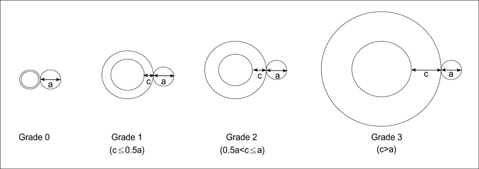
Fig. 2 Schematic diagram depicts four grades of bronchial wall thickening scores. (a) arterial diameter, (c) bronchial wall thickness.
Reference
-
1. Barker AF. Bronchiectasis. N Engl J Med. 2002. 346:1383–1393.
Article2. Naidich DP, McCauley DI, Khouri NF, Stitik FP, Siegelman SS. Computed tomography of bronchiectasis. J Comput Assist Tomogr. 1982. 6:437–444.
Article3. Muller NL, Bergin CJ, Ostrow DN, Nichols DM. Role of computed tomography in the recognition of bronchiectasis. Am J Roentgenol. 1984. 143:971–976.
Article4. Wong-You-Cheong JJ, Leahy BC, Taylor PM, Church SE. Airways obstruction and bronchiectasis: correlation with duration of symptoms and extent of bronchiectasis on computed tomography. Clin Radiol. 1992. 45:256–259.
Article5. Hansell DM, Wells AU, Rubens MB, Cole PJ. Bronchiectasis: funtional significance of areas of decreased attenuation at expiratory CT. Radiology. 1994. 193:369–374.6. Smith IE, Jurriaans E, Diederich S, Ali N, Shneerson JM, Flower CD. Chronic sputum production: correlations between clinical features and findings on high resolution computed tomographic scanning of the chest. Thorax. 1996. 51:914–918.
Article7. Swartz MN. Fishman AP, Elias JA, Grippi MA, Kaiser LR, Senior RM, editors. Bronchiectasis. Fishman's pulmonary diseases and disorders. 1998. 3rd ed. New York: McGraw Hill;2045–2070.8. Cochrane GM, Webber BA, Clarke SW. Effects of sputum on pulmonary function. Br Med J. 1977. 2:1181–1183.
Article9. Perry KMA, King DS. Bronchiectasis: a study of prognosis based on a follow-up of 400 patients. Am Rev Tuberc. 1940. 41:531–548.10. Cherniack NS, Carton RW. Factors associated with respiratory insufficiency in bronchiectasis. Am J Med. 1966. 41:562–571.
Article11. Bhalla M, Turcios N, Aponte V, Jenkins M, Leitman BS, McCauley DI, Naidich DP. Cystic fibrosis: scoring system with thin-section CT. Radiology. 1991. 179:783–788.
Article12. Ip M, Lauder IJ, Wong WY, Lam WK, So SY. Multivariate analysis of factors affecting pulmonary function in bronchiectasis. Respiration. 1993. 60:45–50.
Article13. Ooi GC, Khong PL, Chan-Yeung M, Ho JCM, Chan PKS, Lee JCK, Lam WK, Tsang KWT. High-resolution CT quantification of bronchiectasis: clinical and functional correlation. Radiology. 2002. 225:663–672.
Article14. Roberts HR, Wells AU, Milne DG, Rubens MB, Kolbe J, Cole PJ, Hansell DM. Airflow obstruction in bronchiectasis: correlation between computed tomography features and pulmonary function tests. Thorax. 2000. 55:198–204.
Article15. Miszkiel KA, Wells AU, Rubens MB, Cole PJ, Hansell DM. Effects of airway infection by Pseudomonas aeruginosa: a computed tomographic study. Thorax. 1997. 52:260–264.
Article16. Webb WR, Muller NL, Naidich DP. Webb WR, Muller NL, Naidich DP, editors. Diseases characterized primarily by decreased lung opacity, including cystic abnormalities, emphysema, and bronchiectasis. High-resolution CT of the lung. 1996. 2nd ed. Philadelphia: Lippincott-Raven Publishers;234–241.17. American Thoracic Society. Standardization of spirometry, 1994 update. Am J Respir Crit Care Med. 1995. 152:1107–1136.18. Quanjer PH, Tammeling GJ, Cotes JE, Pedersen OF, Peslin R, Yernault JC. Lung volumes and forced ventilatory flows. Official statement of the European Respiratory Society. Eur Respir J Suppl. 1993. 16:5–40.19. Morris JF. Spirometry in the evaluation of pulmonary function. West J Med. 1976. 125:110–118.20. Reid LM. Reduction in bronchial subdivision in bronchiectasis. Thorax. 1950. 5:233–247.
Article21. Ellis DA, Thornley PE, Wightman AJ, Walker M, Chalmers J, Crofton JW. Present outlook in bronchiectasis: clinical and social study and review of factors influencing prognosis. Thorax. 1981. 36:659–664.
Article22. Osborne D, Vock P, Godwin JD, Silverman PM. CT identification of bronchopulmonary segments: 50 normal subjects. Am J Roentgenol. 1984. 142:47–52.
Article23. O'Riordan T, Wanner A. Baum GL, editor. Bronchiectasis. Textbook of pulmonary diseases. 1998. 6th ed. Philadelphia: Lippincott-Rave;816–817.24. Loubeyre P, Paret M, Revel D, Wiesendanger T, Brune J. Thin-section CT detection of emphysema associated with bronchiectasis and correlation with pulmonary function tests. Chest. 1996. 109:360–365.
Article25. Bahous J, Cartier A, Pineau L, Bernard C, Ghezzo H, Martin RR, Malo JL. Pulmonary function tests and airway responsiveness to methacholine in chronic bronchiectasis of the adult. Bull Eur Physiopathol Respir. 1984. 20:375–380.26. Dail DH. Dail DH, Hammar SP, editors. Bronchial and transbronchial diseases. Pulmonary pathology. 1994. 2nd ed. New York: Springer-Verlag;95–99.
Article27. Comroe JH Jr, Nadel JA. Screening tests of pulmonary function. N Engl J Med. 1970. 282:1249–1253.
Article28. American Thoracic Society. Lung function test: selection of reference values and interpretative strategies. Am Rev Respir Dis. 1991. 144:1202–1218.29. Lynch DA, Newell J, Hale V, Dyer D, Corkery K, Fox NL, Gerend P, Fick R. Correlation of CT findings with clinical evaluations in 261 patients with symptomatic bronchiectasis. Am J Roentgenol. 1999. 173:53–58.
Article30. Nicotra MB, Rivera M, Dale AM, Shepherd R, Carter R. Clinical, pathophysiologic, and microbiologic characterization of bronchiectasis in an aging cohort. Chest. 1995. 108:955–961.
Article31. Angrill J, Agusti C, de Celis R, Filella X, Rano A, Elena M, De La Bellacasa JP, Xaubet A, Torres A. Bronchial inflammation and colonization in patients with clinically stable bronchiectasis. Am J Respir Crit Care Med. 2001. 164:1628–1632.
Article32. Angrill J, Agusti C, de Celis R, Rano A, Gonzalez J, Sole T, Xaubet A, Rodriguez-Roisin R, Torres A. Bacterial colonization in patients with bronchiectasis: microbiological pattern and risk factors. Thorax. 2002. 57:15–19.33. Evans SA, Turner SM, Bosch BJ, Hardy CC, Woodhead MA. Lung function in bronchiectasis: the influence of Pseudomonas aeruginosa. Eur Respir J. 1996. 9:1601–1604.34. Ho PL, Chan KN, Ip MSM, Lam WK, Ho CS, Yuen KY, Tsang KWT. The effect of Pseudomonas aeruginosa infection on clinical parameters in steady-state bronchiectasis. Chest. 1998. 114:1594–1598.
Article35. Rivera M, Nicotra MB. Pseudomonas aeruginosa mucoid strain. Its significance in adult chest diseases. Am Rev Respir Dis. 1982. 126:833–836.36. Woods DE, Straus DC, Johanson WG Jr, Berry VK, Bass JA. Role of pili in adherence of Pseudomonas aeruginosa to mammalian buccal epithelial cells. Infect Immun. 1980. 29:1146–1151.37. Barghouthi S, Guerdoud LM, Speert DP. Inhibition by dextran of Pseudomonas aeruginosa adherence to epithelial cells. Am J Respir Crit Care Med. 1996. 154:1788–1793.
Article
- Full Text Links
-
- Actions
-
Cited
- CITED
-
- Close
- Share
-
- Similar articles
-
- High-Resolution CT Findings in Swyer-James Syndrome
- High-Resolution CT Appearance of Pulmonary Parenchymal Abnormalities Associated with Bronchiectasis: Correlation with Pulmonary Function Tests
- Marked Recovery From Paraquat-Induced Lung Injury During Long-Term Follow-up
- The Correlation between the Radiological Changes and the Level of Transforming Growth Factor-beta1 in Patients with Pulmonary Tuberculosis
- Imaging Diagnosis of Asbestosis
