The Distribution of Fetal Nuchal Translucency Thickness in Normal Korean Fetuses
- Affiliations
-
- 1Department of Obstetrics and Gynecology, Samsung Cheil Hospital and Women's Healthcare Center, Sungkyunkwan University, School of Medicine, Seoul, Korea. ryu97@samsung.co.kr
- 2Department of Diagnostic Radiology, Samsung Cheil Hospital and Women's Healthcare Center, Sungkyunkwan University, School of Medicine, Seoul, Korea.
- 3Laboratory of Medical Genetics, Samsung Cheil Hospital and Women's Healthcare Center, Sungkyunkwan University, School of Medicine, Seoul, Korea.
- KMID: 1785690
- DOI: http://doi.org/10.3346/jkms.2004.19.1.32
Abstract
- The aim of present study was to establish normative data for the distribution of nuchal translucency (NT) thickness in normal Korean fetuses. The data were collected from pregnant women with singleton pregnancies in whom fetal ultrasound was performed and the fetal NT thickness was measured between 11 and 14 weeks of gestation. Among them, a total of 2,577 fetuses with a known normal outcome were included in this study. The distribution of multiple of median (MoM) values of the NT thickness with crown-rump length (CRL) in 10-mm intervals and the 95th percentile of MoM were calculated with the linear regression method. The present study showed that NT measurements increase with increasing CRL and a false positive rate increases with increasing gestational age. Therefore, a fixed cut-off point through the first trimester was not appropriate and each NT measurement should be examined according to the gestational age. The present study offers normative data of the fetal NT thickness in a Korean population, which can be used as reference for screening chromosomal aberrations or other congenital abnormalities in the first trimester.
MeSH Terms
Figure
-
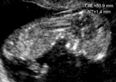
Fig. 1 Measurement of the nuchal translucency (NT) thickness on transvaginal ultrasound scan in 12.0 weeks sized fetus.
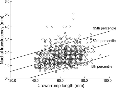
Fig. 2 The distribution of nuchal translucency (NT) thickness in normal fetuses with crown-rump length.
Cited by 2 articles
-
Threshold of Nuchal Translucency for the Detection of Chromosomal Aberration: Comparison of Different Cut-offs
Min-Hyoung Kim, Su-Hyun Park, Hye-Jin Cho, June-Seek Choi, Joo-Oh Kim, Hyun-Kyong Ahn, Joong-Sik Shin, Jung-Yeol Han, Moon-Young Kim, Jae-Hyug Yang
J Korean Med Sci. 2006;21(1):11-14. doi: 10.3346/jkms.2006.21.1.11.Chromosomal Abnormalities in Korean Fetuses with Nuchal Translucency above the 99th Percentile
Dong Wook Kwak, Hyeyeon Boo, Eun Hye Chang, Hyun Mee Ryu, You Jung Han, Jin Hoon Chung, Moon Young Kim, Eun Jung Yang, Hye Ji Yoo, Jin Woo Kim
Perinatology. 2019;30(2):78-82. doi: 10.14734/PN.2019.30.2.78.
Reference
-
1. Nicolaides KH, Brizot ML, Snijders RJ. Fetal nuchal translucency: ultrasound screening for fetal trisomy in the first trimester of pregnancy. Br J Obstet Gynaecol. 1994. 101:782–786.
Article2. Pandya PP, Snijders RJ, Johnson SP, De Lourdes Brizot M, Nicolaides KH. Screening for fetal trisomies by maternal age and fetal nuchal translucency thickness at 10 to14 weeks of gestation. Br J Obstet Gynaecol. 1995. 102:957–962.3. Pandya PP, Kondylios A, Hilbert L, Snijders RJ, Nicolaides KH. Chromosomal defects and outcome in 1015 fetuses with increased nuchal translucency. Ultrasound Obstet Gynecol. 1995. 5:15–19.
Article4. Hyett J, Perdu M, Sharland G, Snijders R, Nicolaides KH. Using fetal nuchal translucency to screen for major congenital cardiac defects at 10-14 weeks of gestation: population based cohort study. Br Med J. 1999. 318:81–85.5. Hafner E, Schuchter K, Liebhart E, Philipp K. Results of routine fetal nuchal translucency measurement at weeks 10-13 in 4233 unselected pregnant women. Prenat Diagn. 1998. 18:29–34.
Article6. Spencer K, Spencer CE, Power M, Moakes A, Nicolaides KH. One stop clinic for assessment of risk for fetal anomalies: a report of the first year of prospective screening for chromosomal anomalies in the first trimester. BJOG. 2000. 107:1271–1275.
Article7. Watt HC, Wald NJ, Smith D, Kennard A, Densem J. Effect of allowing for ethnic group in prenatal screening for Down's syndrome. Prenat Diagn. 1996. 16:691–698.
Article8. Spencer K, Ong CY, Liao AW, Nicolaides KH. The influence of ethnic origin on first trimester biochemical markers of chromosomal abnormalities. Prenat Diagn. 2000. 20:491–494.
Article9. Thilaganathan B, Khare M, Williams B, Wathen NC. Influence of ethnic origin on nuchal translucency screening for Down's syndrome. Ultrasound Obstet Gynecol. 1998. 12:112–114.
Article10. Park JW, Lee WK. Changes of nuchal translucency in early normal fetuses. Korean J Obstet Gynecol. 2000. 43:998–1001.11. Lee K, Cha DH, Park SP, Park HJ. Nuchal translucency measurement in normal fetuses at 10-14 weeks of gestation I. Korean J Obstet Gynecol. 2000. 43:1822–1827.12. Lee K, Cha DH, Kim JH, Seo SK, Lee DW, Cho SH, Kwon JY. Nuchal translucency measurement in normal Korean fetuses at 10-14 weeks of gestation (II). Korean J Obstet Gynecol. 2003. 46:522–527.13. Hadlock FP, Shah YP, Kanon DJ, Lindsey JV. Fetal crown-rump length: Reevaluation of relation to menstrual age (5-18 weeks) with high-resolutin real-time US. Radiology. 1992. 182:501–505.14. Ville Y, Lalondrelle C, Doumerc S, Daffos F, Frydman R, Oury JF, Dumez Y. First trimester diagnosis of nuchal anomalies: significance and fetal outcome. Ultrasound Obstet Gynecol. 1992. 2:314–316.15. Whitlow BJ, Chatzipapas IK, Economides DL. The effect of fetal neck position on nuchal translucency measurement. Br J Obstet Gynaecol. 1998. 105:872–876.
Article16. Snijders RJ, Noble P, Sebire N, Souka A, Nicolaides KH. UK multicentre project on assessment of risk of trisomy 21 by maternal age and fetal nuchal-translucency thickness at 10-14 weeks of gestation. Lancet. 1998. 352:343–346.
Article17. Scott F, Boogert A, Sinosich M, Anderson J. Establishment and application of a normal range for nuchal translucency across the first trimester. Prenat Diagn. 1996. 16:629–634.
Article18. Taipale P, Hiilesmaa V, Salonen R, Ylostalo P. Increased nuchal translucency as a marker for fetal chromosomal defects. N Engl J Med. 1997. 337:1654–1658.
Article19. Schuchter K, Wald N, Hackshaw AK, Hafner E, Liebhart E. The distribution of nuchal translucency at 10-13 weeks of pregnancy. Prenat Diagn. 1998. 18:281–286.
Article20. Wald NJ, Cuckle HS, Densem JW, Nanchahal K, Royston P, Chard T, Haddow JE, Knight GJ, Palomaki GE, Canick JA. Maternal serum screening for Down's syndrome in early pregnancy. Br Med J. 1988. 297:883–887.
Article21. Snijders RJ, Johnson S, Sebire NJ, Noble PL, Nicolaides KH. First-trimester ultrasound screening for chromosomal defects. Ultrasound Obstet Gynecol. 1996. 7:216–226.
Article22. Kim MY, Ryu HM, Kim ES, Han HW, Yang JH, Yoo SJ, Lee YH, Han JR, Lee KS. The value of increased nuchal translucency (NT) for the prediction of abnormal pregnancy outcome. Korean J Perinatol. 1998. 9:363–374.23. Lee JY, Choi KH, Park CW, Yun TS, Park CJ, Jang PR, Park YS. Fetal nuchal translucency measurement for detection of chromosomal abnormalities in the first trimester of high risk pregnancy. Korean J Obstet Gynecol. 1998. 41:2739–2742.24. Kim SJ, Kim CM, Min BS, Sohn WS, Kang JB, Jang PR. The efficacy of nuchal translucency with free beta-hCG, PAPP-A as a screening test for detection of chromosomal anomaly in the first trimester of pregnancy. Korean J Obstet Gynecol. 2001. 44:1091–1096.25. Jou HJ, Wu SC, Li TC, Hsu HC, Tzeng CY, Hsieh FJ. Relationship between fetal nuchal translucency and crown-rump length in an Asian population. Ultrasound Obstet Gynecol. 2001. 17:111–114.
Article26. Chen M, Lam YH, Tang MH, Lee CP, Sin SY, Tang R, Wong HS, Wong SF. The effect of ethnic origin on nuchal translucency at 10-14 weeks of gestation. Prenat Diagn. 2002. 22:576–578.
Article27. Cicero S, Curcio P, Papageorghiou A, Sonek J, Nicolaides K. Absence of nasal bone in fetuses with trisomy 21 at 11-14 weeks of gestation: an observational study. Lancet. 2001. 358:1665–1667.
Article28. Cicero S, Bindra R, Rembouskos G, Spencer K, Nicolaides KH. Integrated ultrasound and biochemical screening for trisomy 21 using fetal nuchal translucency, absent fetal nasal bone, free β-hCG and PAPP-A at 11 to 14 weeks. Prenat Diagn. 2003. 23:306–310.
Article
- Full Text Links
-
- Actions
-
Cited
- CITED
-
- Close
- Share
-
- Similar articles
-
- Changes of Nuchal Translucency in Early Normal Fetuses
- Measurements of Fetal Nuchal Skinfold Thickness in Normal Fetuses during Second Trimester
- Prenatal chromosomal microarray analysis of fetus with increased nuchal translucency
- Nuchal Translucency Measurement in Normal Fetuses at 10 - 14 Weeks of Gestation I
- First trimester screening for trisomy 18 by a combination of nuchal translucency thickness and epigenetic marker level
