Korean J Radiol.
2010 Apr;11(2):231-233. 10.3348/kjr.2010.11.2.231.
Perforated Sigmoid Colon Cancer within an Irreducible Inguinal Hernia: a Case Report
- Affiliations
-
- 1Department of Radiology, Tri-Service General Hospital, National Defense Medical Center, China. chougo2002@yahoo.com.tw
- 2Division of Genitourinary Surgery, Department of Surgery, Tri-Service General Hospital, National Defense Medical Center, China.
- 3Department of Emergency Medicine, Tri-Service General Hospital, National Defense Medical Center, China.
- KMID: 1783200
- DOI: http://doi.org/10.3348/kjr.2010.11.2.231
Abstract
- A perforated sigmoid colon cancer within an inguinal hernia is extremely rare. This unexpected finding is usually discovered during surgery and causes an unavoidable septic evolution. Here, we describe the case of an 84-year-old man who presented with fever, abdominal distension, and a painful, enlarged, left scrotum. A CT showed a left, incarcerated, inguinal hernia containing a perforated sigmoid adenocarcinoma (which was confirmed by histopathology). The possibility of an irreducible inguinal hernia in association with perforated sigmoid colon cancer should be considered in the array of diagnoses. A pre-operative CT scan would be helpful in facilitating an accurate diagnosis.
MeSH Terms
-
Adenocarcinoma/complications/*radiography/surgery
Aged, 80 and over
Colon, Sigmoid/radiography/surgery
Diagnosis, Differential
Fatal Outcome
Fever/etiology
Hernia, Inguinal/complications/*radiography/surgery
Humans
Intestinal Perforation/complications/*radiography/surgery
Male
Pain/etiology
Shock, Septic/complications
Sigmoid Neoplasms/complications/*radiography/surgery
Tomography, X-Ray Computed
Figure
-
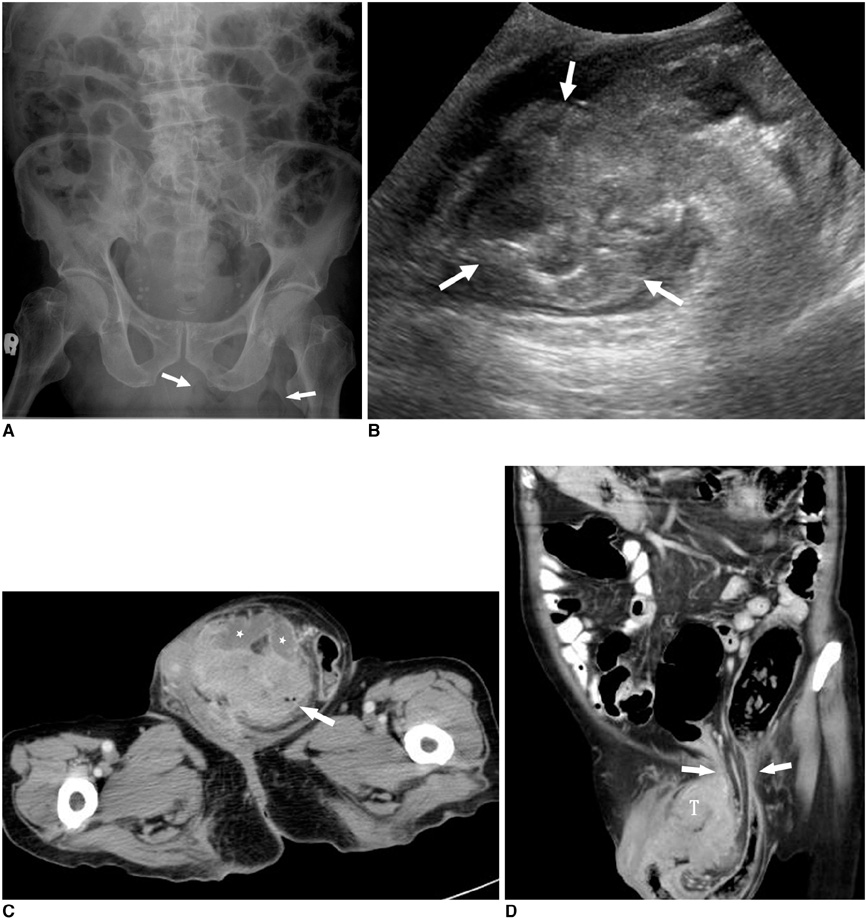
Fig. 1 Colonic perforation within irreducible inguinal hernia. A. Plain abdominal radiograph shows increased soft tissue density including suspicious bowel-gas (arrows) in left inguinal region with ileus of intraabdominal bowel loops. B. Ultrasound of scrotum shows large heterogeneously echogenic mass (arrows) in left scrotum with little fluid collection. C. Axial CT image shows tumor-like mass of sigmoid colon (arrow) within left scrotal sac, which is surrounded by multiple abscesses (asterisks). D. Reformatted coronal CT image shows sigmoid tumor (T) herniating into left scrotal sac through inguinal canal (arrows) and causing dilatation of descending colon. No evidence of peritoneal contamination is observed.
Reference
-
1. Boormans JL, Hesp WL, Teune TM, Plaisier PW. Carcinoma of the sigmoid presenting as a right inguinal hernia. Hernia. 2006. 10:93–96.2. Slater R, Amatya U, Shorthouse AJ. Colonic carcinoma presenting as strangulated inguinal hernia: report of two cases and review of the literature. Tech Coloproctol. 2008. 12:255–258.3. Tan GY, Guy RJ, Eu KW. Obstructing sigmoid cancer with local invasion in an incarcerated inguinal hernia. ANZ J Surg. 2003. 73:80–82.4. Kouraklis G, Kouskos E, Glinavou A, Raftopoulos J, Karatzas G. Perforated carcinoma of the sigmoid colon in an incarcerated inguinal hernia: report of a case. Surg Today. 2003. 33:707–708.5. Hale DA, Solla JA. Complete colonic obstruction caused by a sigmoid colon cancer incarcerated in an inguinal hernia sac. South Med J. 1991. 84:1280–1281.6. Sakorafas GH, Peros G. Obstructing sigmoid cancer in a patient with a large, tender, non-reducible inguinal hernia: the obvious diagnosis is not always the correct one. Eur J Cancer Care (Engl). 2008. 17:72–73.7. Mandava N, Kumar S, Pizzi WF, Aprile IJ. Perforated colorectal carcinomas. Am J Surg. 1996. 172:236–238.8. Baskin LS, Carroll PR, Cattolica EV, McAninch JW. Necrotising soft tissue infections of the perineum and genitalia. Bacteriology, treatment and risk assessment. Br J Urol. 1990. 65:524–529.
- Full Text Links
-
- Actions
-
Cited
- CITED
-
- Close
- Share
-
- Similar articles
-
- Inguinal hernia containing the uterus and both adnexa in a full-term infant
- Incarcerated Inguinal Hernia in Children
- Giant Transverse Colon Diverticulitis Presenting as Indirect Right Inguinal Hernia Strangulation
- Incarcerated internal hernia within a huge irreducible parastomal hernia with intestinal obstruction: a rare case report of "hernia within hernia"
- An Adult Right-sided Bochdalek Hernia Accompanied with Hepatic Hypoplasia and Inguinal Hernia
