Variations of the Superficial Brachial Artery in Korean Cadavers
- Affiliations
-
- 1Department of Anatomy, Yonsei University College of Medicine, Seoul, Korea. leehy@yumc.yonsei.ac.kr
- KMID: 1783077
- DOI: http://doi.org/10.3346/jkms.2008.23.5.884
Abstract
- The superficial brachial artery (SBA), a branch of the axillary artery, is one of the most common arterial variations in this area. While it is more vulnerable to accidental arterial injection or injury, it could be useful for the nourishment of a medial arm skin free flap. To analyze the relationship between the SBA of axillary origin and segmental variation of the axillary artery, we dissected 304 arms of Korean cadavers. We found an SBA of axillary origin in 12.2% of cadaveric arms. Unilateral occurrence was detected in 16 cadavers and bilateral in 10. SBAs gave rise to radial and ulnar arteries in the cubital fossa (8.9%), continued in the forearm as the radial artery (2.3%), or ended in the upper arm (1.0%). The SBA ended as ulnar artery was not found in any of the cadavers. The bifurcation of the SBA into the radial and ulnar arteries, presence of an SBA that ends in the upper arm, and the lack of continuation as the ulnar artery are characteristics of SBAs in Korean cadavers.
Keyword
MeSH Terms
Figure
-
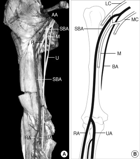
Fig. 1 Photograph (A) and illustration (B) of type I SBA. The SBA bifurcated into the radial and the ulnar arteries after giving muscular branches to the biceps brachii and brachialis muscles. AA, axillary artery; LC, lateral cord; MC, medial cord; M, median nerve; U, ulnar nerve; R, radial nerve; SBA, superficial brachial artery; RA, radial artery; UA, ulnar artery.

Fig. 2 Type Ia, Ib, and Ic superficial brachial arteries. Type Ia (A): the SBA emerged proximal to the stem of the subscapular artery, anterior humeral circumflex artery, and posterior humeral circumflex artery. Type Ib (B): the SBA emerged from the axillary artery distal to the stem of the subscapular artery in the second part, proximal to the stems of the circumflex arteries. Type Ic (C): the SBA emerged distal to the thoracodorsal artery from the second part of the axillary artery, proximal to the circumflex scapular artery appearing from the third part. AA, axillary artery; SBA, superficial brachial artery; SA, subscapular artery; AH, anterior humeral circumflex artery; PH, posterior humeral circumflex artery; TA, thoracodorsal artery; CA, circumflex scapular artery; LC, lateral cord; MC, medial cord; M, median nerve.

Fig. 3 Photograph (A) and illustration (B) of type II SBA. After giving muscular branches to the biceps brachii and brachialis muscles, the SBA continued as the radial artery in the forearm. AA, axillary artery; BA, brachial artery; LC, lateral cord; MC, medial cord; M, median nerve; SBA, superficial brachial artery; RA, radial artery; UA, ulnar artery.

Fig. 4 Photograph (A) and illustration (B) of type III SBA. The slender SBA supplied the arm musculature and ended in the upper arm. AA, axillary artery; BA, brachial artery; LC, lateral cord; MC, medial cord; M, median nerve; U, ulnar nerve; SBA, superficial brachial artery; RA, radial artery; UA, ulnar artery.
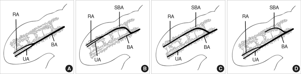
Fig. 5 The development of the brachial artery and the superficial brachial artery in embryonic upper limb. (A) Absent superficial brachial artery. (B) Type I superficial brachial artery. (C) Type II superficial brachial artery. (D) Type III superficial brachial artery. During the limb development, the survival or regression of the arteries are decided by hemodynamic dominance of primitive arterial networks. BA, brachial artery; SBA, superficial brachial artery; RA, radial artery; UA, ulnar artery.
Cited by 2 articles
-
Morphological Assessment of Cadaveric Radial, Brachial and Subclavian Arteries: A Neurointerventional Approach
Ali Yilmaz, Ayca Ozkul, Dong Seong Shin, Soo-Bin Im, Seok-Mann Yoon, Bum-Tae Kim
J Korean Neurosurg Soc. 2015;58(6):499-503. doi: 10.3340/jkns.2015.58.6.499.Study of course and termination of brachial artery by dissection and computed tomography angiography methods with clinical importance
Hemamalini Shetty, Vikram Patil, Najma Mobin, Manjunatha Hanasoge Narayana Gowda, Vinutha Shanubhoganahalli Puttamallappa, Ravishankar Mathada Vamadevaiah, Pushpalatha Kunjappagounder
Anat Cell Biol. 2022;55(3):284-293. doi: 10.5115/acb.22.053.
Reference
-
1. Adachi B. Das Arteriensystem der Japaner. 1928. Vol. 1. Kyoto: Maruzen Press;196–290.2. McCormack LJ, Cauldwell EW, Anson BJ. Brachial and antebrachial arterial patterns; a study of 750 extremities. Surg Gynecol Obstet. 1953. 96:43–54.3. Keen JA. A study of the arterial variations in the limbs, with special reference to symmetry of vascular patterns. Am J Anat. 1961. 108:245–261.
Article4. Fuss FK, Matula CW, Tschabitscher M. Die Arteria brachialis superficialis. Anat Anz. 1985. 160:285–294.5. Anagnostopoulou S, Venieratos D. An unusual branching pattern of the superficial brachial artery accompanied by an ulnar nerve with two roots. J Anat. 1999. 195:471–476.
Article6. Yoshinaga K, Tanii I, Kodama K. Superficial brachial artery crossing over the ulnar and median nerves from posterior to anterior: embryological significance. Anat Sci Int. 2003. 78:177–180.
Article7. Bataineh ZM, Al-Hussain SM, Moqattash ST. Complex neurovascular variation in one upper limb. Ital J Anat Embryol. 2007. 112:37–44.8. D'Costa S, Shenoy BM, Narayana K. The incidence of a superficial arterial pattern in the human upper extremities. Folia Morphol (Warsz). 2004. 63:459–463.9. Kawashima T, Yoshitomi S, Sasaki H. Anatomical relationship between the superficial brachial arteries and the brachial plexus in humans, and their morphological significance. Folia Morphol (Warsz). 2004. 63:465–471.10. Coskun N, Sarikcioglu L, Donmez BO, Sindel M. Arterial, neural and muscular variations in the upper limb. Folia Morphol (Warsz). 2005. 64:347–352.11. Yalcin B, Kocabiyik N, Yazar F, Kirici Y, Ozan H. Arterial variations of the upper extremities. Anat Sci Int. 2006. 81:62–64.
Article12. Hollinshead WH. Anatomy for surgeons. 1982. vol.3, the back and limbs:3rd ed. Philadelphia: Harper & Row;362.13. Karamürsel S, Bağdatli D, Demir Z, Tüccar E, Celebioğlu S. Use of medial arm skin as a free flap. Plast Reconstr Surg. 2005. 115:2025–2031.14. Rodríguez-Baeza A, Nebot J, Ferreira B, Reina F, Pérez J, Sañudo JR, Roig M. An anatomical study and ontogenetic explanation of 23 cases with variations in the main pattern of the human brachio-antebrachial arteries. J Anat. 1995. 187:473–479.15. Singer E. Human brachial plexus united into a single cord description and interpretation. Anat Rec. 1933. 55:411–419.
Article16. Melling M, Wilde J, Schnallinger M, Karimian-Teherani D, Behnam M, Firbas W. Rare variant of the brachial artery: superficial lateral inferior type VII EAB. Clin Anat. 2000. 13:216–222.
Article17. Skopakoff C. Uber die Variabilitat der Ab-und Verzweigung der A. brachialis superficialis. Anat Anz. 1959. 106:356–368.
- Full Text Links
-
- Actions
-
Cited
- CITED
-
- Close
- Share
-
- Similar articles
-
- Study of course and termination of brachial artery by dissection and computed tomography angiography methods with clinical importance
- A Case of Superficial Brachial Artery
- Complex Variations in Branching Pattern of the Axillary Artery and Hands with the Persistent Median Artery
- Anatomical variation of median nerve: cadaveric study in brachial plexus
- Superficial brachioulnar artery and its clinical significance
