Study of course and termination of brachial artery by dissection and computed tomography angiography methods with clinical importance
- Affiliations
-
- 1Department of Anatomy, JSS Medical College, JSS AHER, Mysuru, India.
- 2Department of Radiology, JSS Hospital, Mysuru, India.
- KMID: 2533352
- DOI: http://doi.org/10.5115/acb.22.053
Abstract
- The Brachial artery is a continuation of the axillary artery, from the inferior border of the tendon of teres major to the neck of the radius, terminating into radial and ulnar arteries just a cm distal to the elbow joint. Unlike veins, variations in the arteries are comparatively less common. Anatomical variations of the brachial artery occur in almost 20% of the cases and are commonly found during routine dissection or clinical practice. To observe the variations in the course and termination of brachial artery by dissection and computed tomography (CT) angiography methods. The present study was conducted on 40 upper limbs each in the department of Anatomy & Radiology of JSS Medical College and Hospital, Mysuru. The brachial artery was traced from origin to termination and variations were noted and photographed. Patients who were undergoing CT angiography of the upper limbs in JSS Hospital were included in the study. Variations noted and compared with the dissection method. In the present study, normal patterns of the brachial arterial course and termination were observed in 31 specimens. The remaining 9 specimens showed variant course and termination in the brachial artery like an unusually tortuous superficial brachial artery, superficial brachio-ulnar artery and brachio-radial artery. CT angiography showed 6 variations and a tortuous brachial artery. A detailed description of the vascular pattern of upper limbs especially variations in their origin and termination is of extreme importance in clinical practice. The knowledge of these variations is important for catheterization, graft harvesting, arteriovenous fistula creation, shunt application and astrup examination.
Keyword
Figure
-

Fig. 1 Right upper limb showing unusually TSBA. TSBA, tortuous superficial brachial artery; MN, median nerve; RA, radial artery; UA, ulnar artery.

Fig. 2 Left upper limb showing TSBA. TSBA, tortuous superficial brachial artery; RA, radial artery; UA, ulnar artery; MN, median nerve.
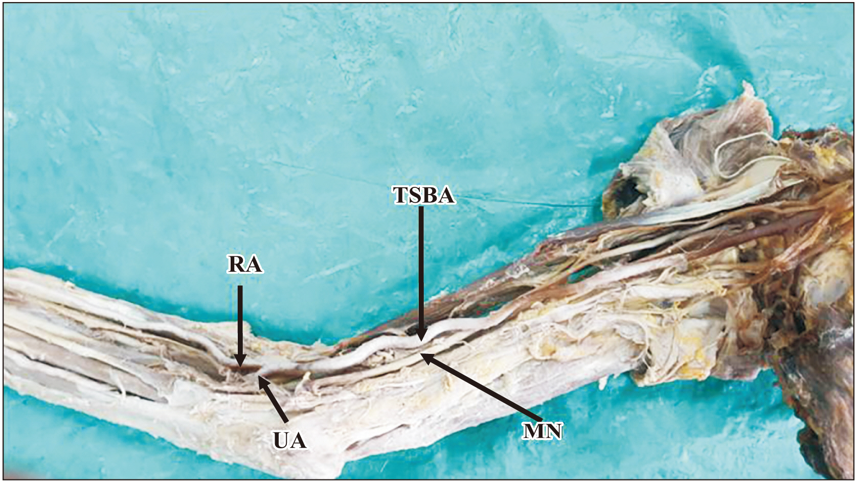
Fig. 3 Right upper limb showing TSBA. TSBA, tortuous superficial brachial artery; RA, radial artery, UA, ulnar artery; MN, median nerve.
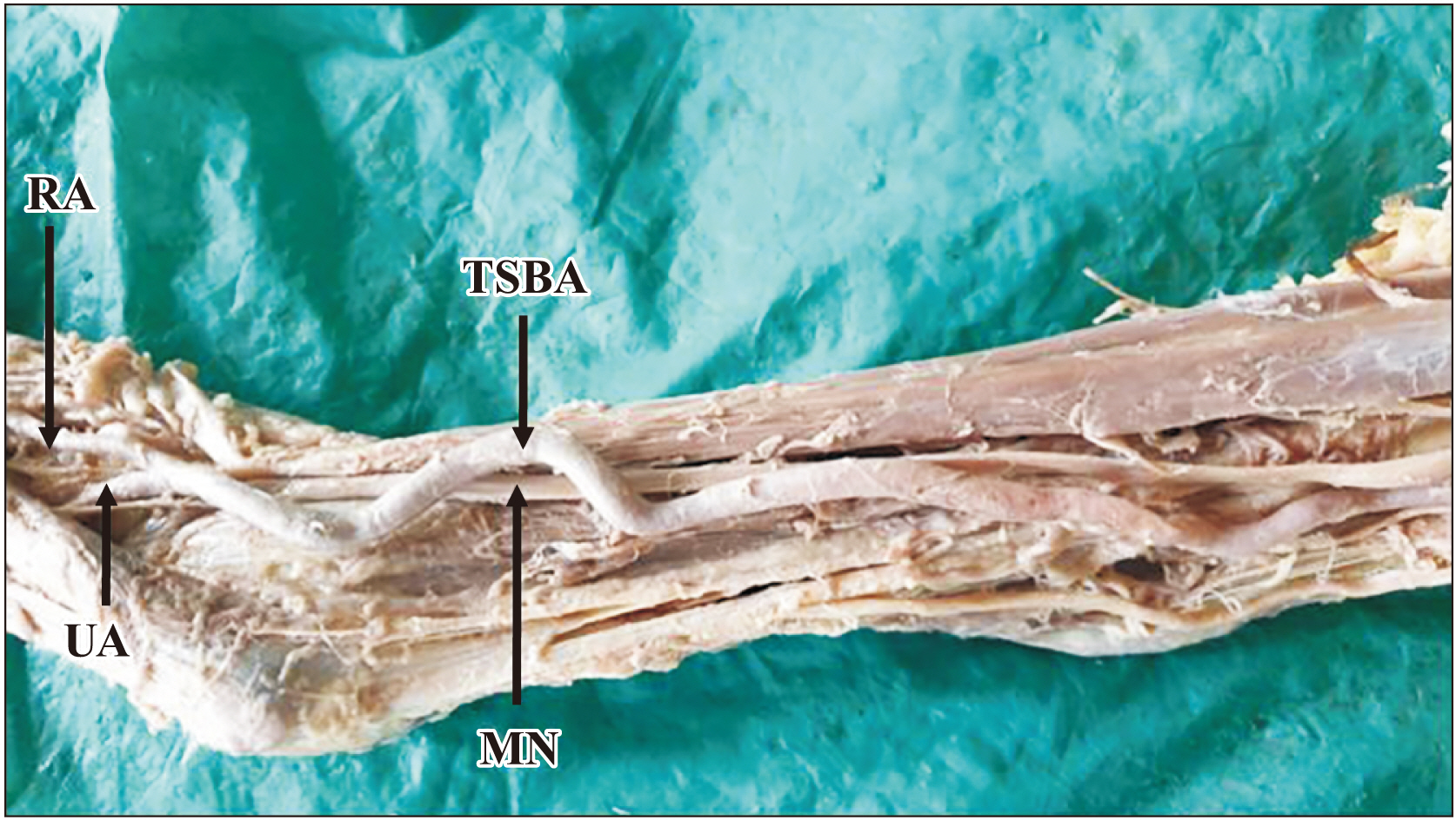
Fig. 4 Right upper limb showing TSBA. TSBA, tortuous superficial brachial artery; RA, radial artery; UA, ulnar artery; MN, median nerve.
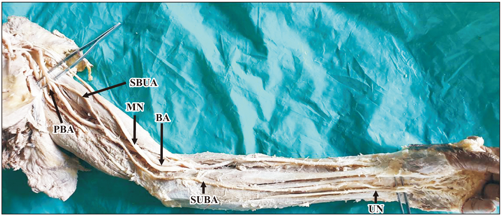
Fig. 5 Left upper limb showing SBUA or high origin of ulnar artery. SBUA, superficial brachio-radial artery; BA, brachial artery; MN, median nerve; PBA, profunda brachii artery, UN, ulnar nerve.
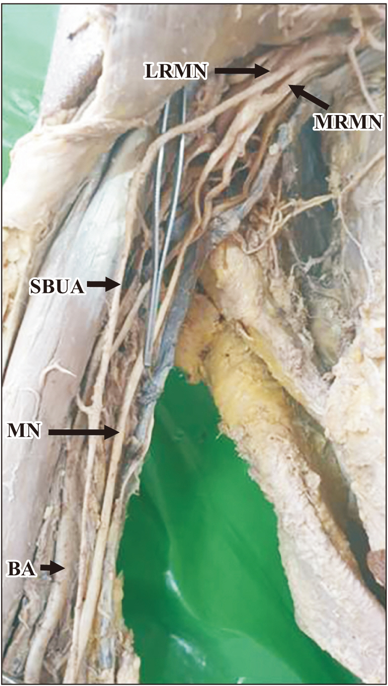
Fig. 6 Right upper limb showing high origin of ulnar artery from axillary artery. LRMN, lateral root of the median nerve; MRMN, medial root of the median nerve; SBUA, superficial brachio-radial artery; MN, median nerve; BA, brachial artery.
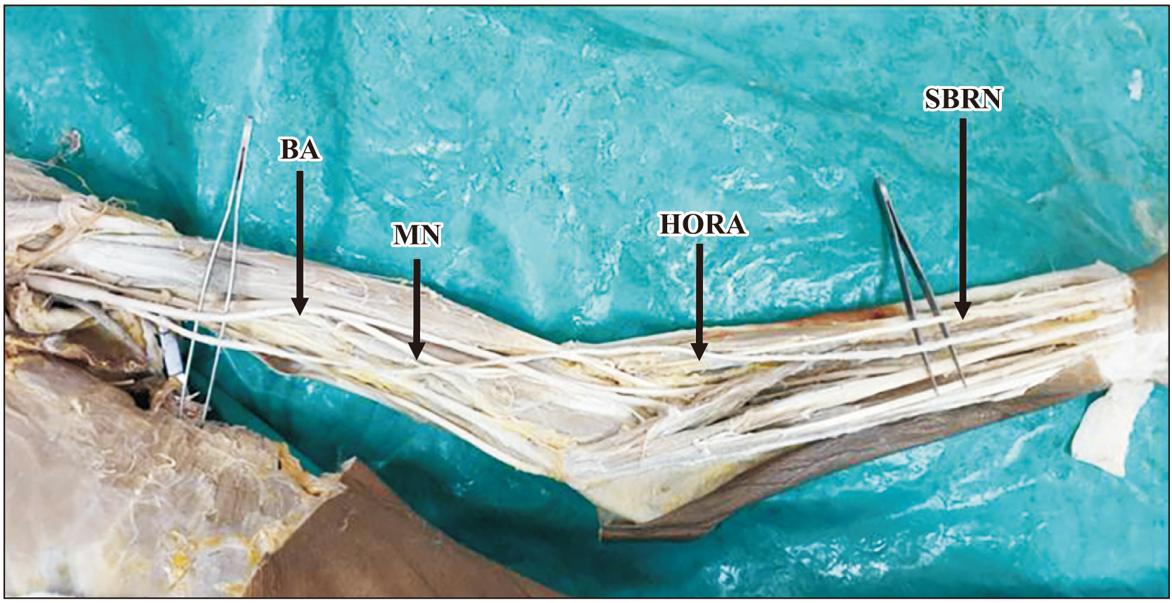
Fig. 7 Left upper limb showing brachio-radial artery or high origin of radial artery. SBRN, superficial branch of radial nerve; HORA, high origin of radial artery; MN, median nerve; BA, brachial artery.

Fig. 8 Right upper limb showing brachio-radial artery or HORA. HORA, high origin of radial artery; MN, median nerve; BA, brachial artery.
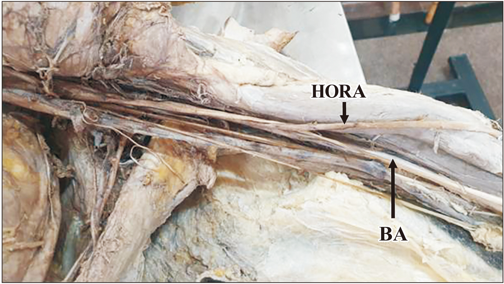
Fig. 9 Left upper limb showing brachio-radial artery or HORA. HORA, high origin of radial artery; BA, brachial artery.
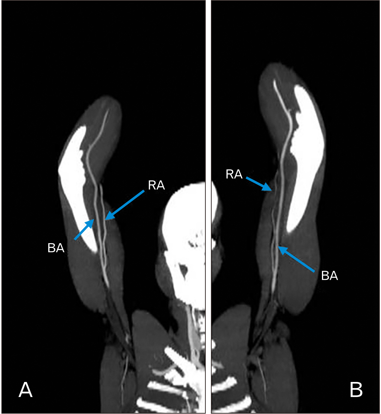
Fig. 10 Computed tomography angiography images showing brachio-radial artery and tortuous brachial artery. (A) High origin of radial artery, (B) high origin of radial artery. RA, radial artery; BA, brachial artery.
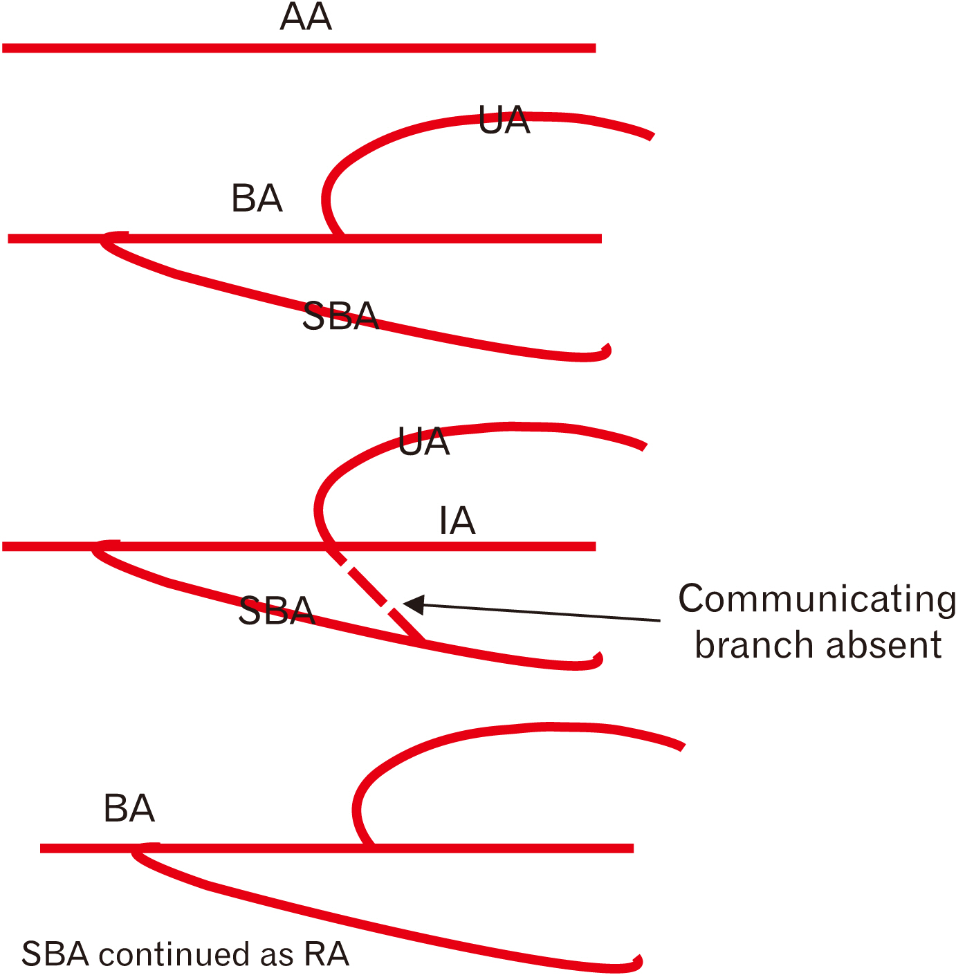
Fig. 11 Schematic representation of developmental basis for SBA that continues as radial artery. AA, axis artery; UA, ulnar artery; BA, brachial artery; SBA, superficial brachial artery; IA, interosseous artery; RA, radial artery.

Fig. 12 Schematic representation of developmental basis for SBUA. AA, axis artery; SBUA, superficial brachio-ulnar artery; BA, brachial artery; SBA, superficial brachial artery; IA, interosseous artery; RA, radial artery.
Reference
-
References
1. Standring S. 2008. Gray's anatomy: the anatomical basis of clinical practice. 40th ed. Churchill Livingstone;Edinburgh: p. 905–6.2. Rodríguez-Niedenführ M, Vázquez T, Nearn L, Ferreira B, Parkin I, Sañudo JR. 2001; Variations of the arterial pattern in the upper limb revisited: a morphological and statistical study, with a review of the literature. J Anat. 199(Pt 5):547–66. DOI: 10.1046/j.1469-7580.2001.19950547.x. PMID: 11760886. PMCID: PMC1468366.
Article3. Singer E. 1933; Embryological pattern persisting in the arteries of the arm. Anat Rec. 55:403–9. DOI: 10.1002/ar.1090550407.
Article4. Han HC. 2012; Twisted blood vessels: symptoms, etiology and biomechanical mechanisms. J Vasc Res. 49:185–97. DOI: 10.1159/000335123. PMID: 22433458. PMCID: PMC3369246.
Article5. Weibel J, Fields WS. 1965; Tortuosity, coiling, and kinking of the internal carotid artery. I. Etiology and radiographic anatomy. Neurology. 15:7–18. DOI: 10.1212/WNL.15.1.7. PMID: 14257832.
Article6. Ertugrul A. 1967; Diffuse tortuosity and lengthening of the arteries. Circulation. 36:400–7. DOI: 10.1161/01.CIR.36.3.400. PMID: 6033167.
Article7. Ashwini C, Vasantha K. 2014; An unusual tortuous brachial artery and its branches: histological basis and its clinical perspective. Int J Life Sci Biotechnol Pharm Res. 3:121–4.8. Dobrin PB, Schwarcz TH, Baker WH. 1988; Mechanisms of arterial and aneurysmal tortuosity. Surgery. 104:568–71. PMID: 3413685.9. Du Toit DF, Sunshine M, Knott-Craig C, Laker L. 1985; Gangrene of the hand and forearm after inadvertent intra-arterial injection of pyrazole. A case report. S Afr Med J. 68:491–2. PMID: 4049165.10. Handlogten KS, Wilson GA, Clifford L, Nuttall GA, Kor DJ. 2014; Brachial artery catheterization: an assessment of use patterns and associated complications. Anesth Analg. 118:288–95. DOI: 10.1213/ANE.0000000000000082. PMID: 24445630.11. Tubbs R, Shoja MM, Loukas M. 2016. Bergman's comprehensive encyclopedia of human anatomic variation. John Wiley & Sons;New York: DOI: 10.1002/9781118430309.12. Segal R, Machiraju U, Larkins M. 1992; Tortuous peripheral arteries: a cause of focal neuropathy. Case report. J Neurosurg. 76:701–4. DOI: 10.3171/jns.1992.76.4.0701. PMID: 1545266.13. Nkomozepi P, Xhakaza N, Swanepoel E. 2017; Superficial brachial artery: a possible cause for idiopathic median nerve entrapment neuropathy. Folia Morphol (Warsz). 76:527–31. DOI: 10.5603/FM.a2017.0013. PMID: 28198531.
Article14. Gan HW, Yip HK, Wu CJ. 2010; Brachial approach for coronary angiography and intervention: totally obsolete, or a feasible alternative when radial access is not possible? Ann Acad Med Singap. 39:368–73. PMID: 20535426.15. Alvarez-Tostado JA, Moise MA, Bena JF, Pavkov ML, Greenberg RK, Clair DG, Kashyap VS. 2009; The brachial artery: a critical access for endovascular procedures. J Vasc Surg. 49:378–85. discussion 385DOI: 10.1016/j.jvs.2008.09.017. PMID: 19028057.
Article16. Panagouli E, Anagnostopoulou S, Venieratos D. 2014; Bilateral asymmetry of the highly bifurcated brachial artery variation. Rom J Morphol Embryol. 55:469–72. PMID: 24970004.17. Cherukupalli C, Dwivedi A, Dayal R. 2007; High bifurcation of brachial artery with acute arterial insufficiency: a case report. Vasc Endovascular Surg. 41:572–4. DOI: 10.1177/1538574407305798. PMID: 18166644.
Article18. Tsoucalas G, Eleftheriou A, Panagouli E. 2020; High bifurcation of the brachial artery: an embryological overview. Cureus. 12:e7097. DOI: 10.7759/cureus.7097. PMID: 32231893. PMCID: PMC7098415.
Article19. Yang HJ, Gil YC, Jung WS, Lee HY. 2008; Variations of the superficial brachial artery in Korean cadavers. J Korean Med Sci. 23:884–7. DOI: 10.3346/jkms.2008.23.5.884. PMID: 18955798. PMCID: PMC2580020.
Article20. Lioupis C, Mistry H, Junghans C, Haughey N, Freedman B, Tyrrell M, Valenti D. 2010; High brachial artery bifurcation is associated with failure of brachio-cephalic autologous arteriovenous fistulae. J Vasc Access. 11:132–7. DOI: 10.1177/112972981001100209. PMID: 20155716.
Article21. Hansdak R, Arora J, Sharma M, Mehta V, Suri RK, Das S. 2015; Unusual branching pattern of brachial artery - embryological basis and clinicoanatomical insight. Clin Ter. 166:65–7. DOI: 10.7417/CT.2015.1817. PMID: 25945432.22. Natsis K, Papadopoulou AL, Paraskevas G, Totlis T, Tsikaras P. 2006; High origin of a superficial ulnar artery arising from the axillary artery: anatomy, embryology, clinical significance and a review of the literature. Folia Morphol (Warsz). 65:400–5. PMID: 17171623.23. Jacquemin G, Lemaire V, Medot M, Fissette J. 2001; Bilateral case of superficial ulnar artery originating from axillary artery. Surg Radiol Anat. 23:139–43. DOI: 10.1007/s00276-001-0139-2. PMID: 11462864.
Article24. Nakatani T, Tanaka S, Mizukami S, Shiraishi Y, Nakamura T. 1996; The superficial ulnar artery originating from the axillary artery. Ann Anat. 178:277–9. DOI: 10.1016/S0940-9602(96)80068-9. PMID: 8712378.
Article25. Adachi B. 1928. Das Arteriensystem der Japaner. Volume 1:Maruzen;Kyoto: p. 285–356. German. DOI: 10.1016/s0940-9602(96)80068-9.26. Bozer C, Ulucam E, Kutoglu T. 2004; A case of originated high superficial ulnar artery. Trakia J Sci. 2:70–3.27. Nakatani T, Tanaka S, Mizukami S. 1998; Superficial ulnar artery originating from the brachial artery and its clinical importance. Surg Radiol Anat. 20:383–5. DOI: 10.1007/BF01630626. PMID: 9894322.
Article28. Chin KJ, Singh K. 2005; The superficial ulnar artery--a potential hazard in patients with difficult venous access. Br J Anaesth. 94:692–3. DOI: 10.1093/bja/aei548. PMID: 15814810.
Article29. Latika A, Rima Dada R. 2005; Superficial ulnar artery- a case report. Indian J Pract Dr. 2:7–8. DOI: 10.18410/jebmh/2017/289. PMID: 26715898. PMCID: PMC4681723.30. Jacob J, Deshpande R, Desai J. 2005; Superficial ulnar artery. Eur J Cardio Thorac Surg. 28:494. DOI: 10.1016/j.ejcts.2005.02.048. PMID: 16111612.
Article31. Porter CJ, Mellow CG. 2001; Anatomically aberrant forearm arteries: an absent radial artery with co-dominant median and ulnar arteries. Br J Plast Surg. 54:727–8. DOI: 10.1054/bjps.2001.3706. PMID: 11728121.
Article32. McWilliams RG, Sodha I. 2000; Doppler ultrasound diagnosis of a superficial ulnar artery. Eur J Ultrasound. 12:155–7. DOI: 10.1016/S0929-8266(00)00106-3. PMID: 11118923.
Article33. Sieger J, Patel L, Sheikh K, Parker E, Sheng M, Sakthi-Velavan S. 2019; Superficial brachioulnar artery and its clinical significance. Anat Cell Biol. 52:333–6. DOI: 10.5115/acb.19.008. PMID: 31598363. PMCID: PMC6773909.
Article34. Rodríguez-Baeza A, Nebot J, Ferreira B, Reina F, Pérez J, Sañudo JR, Roig M. 1995; An anatomical study and ontogenetic explanation of 23 cases with variations in the main pattern of the human brachio-antebrachial arteries. J Anat. 187(Pt 2):473–9. PMID: 7592009. PMCID: PMC1167441.35. Weathersby HT. 1956; Anomalies of brachial and antebrachial arteries of surgical significance. South Med J. 49:46–9. DOI: 10.1097/00007611-195601000-00010. PMID: 13281685.36. Karlsson S, Niechajev IA. 1982; Arterial anatomy of the upper extremity. Acta Radiol Diagn (Stockh). 23:115–21. DOI: 10.1177/028418518202300206. PMID: 7090847.
Article37. Uglietta JP, Kadir S. 1989; Arteriographic study of variant arterial anatomy of the upper extremities. Cardiovasc Intervent Radiol. 12:145–8. DOI: 10.1007/BF02577379. PMID: 2507150.
Article38. Jennings WC, Mallios A, Mushtaq N. 2018; Proximal radial artery arteriovenous fistula for hemodialysis vascular access. J Vasc Surg. 67:244–53. DOI: 10.1016/j.jvs.2017.06.114. PMID: 28912005.
Article39. Keen JA. 1961; A study of the arterial variations in the limbs, with special reference to symmetry of vascular patterns. Am J Anat. 108:245–61. DOI: 10.1002/aja.1001080303. PMID: 14454801.
Article40. Patel T, Shah S, Pancholy S, Rao S, Bertrand OF, Kwan T. 2014; Balloon-assisted tracking: a must-know technique to overcome difficult anatomy during transradial approach. Catheter Cardiovasc Interv. 83:211–20. DOI: 10.1002/ccd.24959. PMID: 23592578.
Article41. Ostojić Z, Bulum J, Ernst A, Strozzi M, Marić-Bešić K. 2015; Frequency of radial artery anatomic variations in patients undergoing transradial heart catheterization. Acta Clin Croat. 54:65–72. PMID: 26058245.42. Anderson CB, Etheredge EE, Harter HR, Codd JE, Graff RJ, Newton WT. 1977; Blood flow measurements in arteriovenous dialysis fistulas. Surgery. 81:459–61. PMID: 847655.43. Berman SS, Gentile AT, Glickman MH, Mills JL, Hurwitz RL, Westerband A, Marek JM, Hunter GC, McEnroe CS, Fogle MA, Stokes GK. 1997; Distal revascularization-interval ligation for limb salvage and maintenance of dialysis access in ischemic steal syndrome. J Vasc Surg. 26:393–402. discussion 402–4. DOI: 10.1016/S0741-5214(97)70032-6. PMID: 9308585.
Article44. Odland MD, Kelly PH, Ney AL, Andersen RC, Bubrick MP. 1991; Management of dialysis-associated steal syndrome complicating upper extremity arteriovenous fistulas: use of intraoperative digital photoplethysmography. Surgery. 110:664–9. discussion 669–70. PMID: 1925955.45. Morsy AH, Kulbaski M, Chen C, Isiklar H, Lumsden AB. 1998; Incidence and characteristics of patients with hand ischemia after a hemodialysis access procedure. J Surg Res. 74:8–10. DOI: 10.1006/jsre.1997.5206. PMID: 9536965.
Article46. Zibari GB, Rohr MS, Landreneau MD, Bridges RM, DeVault GA, Petty FH, Costley KJ, Brown ST, McDonald JC. 1988; Complications from permanent hemodialysis vascular access. Surgery. 104:681–6.47. Zamani P, Kaufman J, Kinlay S. 2009; Ischemic steal syndrome following arm arteriovenous fistula for hemodialysis. Vasc Med. 14:371–6. DOI: 10.1177/1358863X09102293. PMID: 19808723.
Article48. Bidarkotimath S, Avadhani R, Arunachalam K. 2011; Primary pattern of arteries of upper limb with relevance to their variations. Int J Morphol. 29:1422–8. DOI: 10.4067/S0717-95022011000400059.
Article49. Vatsala AR, Rajashekar HV, Angadi AV, Sangam . 2013; Variation in the branching pattern of Brachial artery: a morphological and statistical study. Int J Biol Med Res. 4:2920–3.50. Sonje P, Kanaskar N, Arole V, Shevade S, Awari P, Vatsalaswamy . 2014; Study of variations in the branching pattern of brachial artery. Int J Cur Res Rev. 6:55–60.51. Deepa TK, John Martin K. 2016; An anatomical study of variations in termination of brachial artery, with its embryological basis and clinical significance. Int J Med Res Health Sci. 5:85–9.52. Kaur A, Sharma A, Sharma M. 2017; Variation in branching pattern of brachial artery. Int J Sci Stud. 5:213–7.53. Ojha P, Prakash S. 2019; Variations in branching pattern of brachial artery - a study in cadavers. Int J Sci Res. 8:33–6. DOI: 10.36106/ijsr/1300946.
Article54. Balakrishnan R, Kavya , Sharmada KL. 2020; Arterial variation in the brachial artery and its clinical implications. MedPulse Int J Anat. 13:5–8. DOI: 10.26611/10011312.55. Konarik M, Musil V, Baca V, Kachlik D. 2020; Upper limb principal arteries variations: a cadaveric study with terminological implication. Bosn J Basic Med Sci. 20:502–13. DOI: 10.17305/bjbms.2020.4643. PMID: 32343941. PMCID: PMC7664784. PMID: 2ff53371eadb4f6ebb70390d1b74ab85.
Article56. Celik HH, Görmüs G, Aldur MM, Ozçelik M. 2001; Origin of the radial and ulnar arteries: variations in 81 arteriograms. Morphologie. 85:25–7. PMID: 11534414.
- Full Text Links
-
- Actions
-
Cited
- CITED
-
- Close
- Share
-
- Similar articles
-
- A Case of Spontaneous Coronary Artery Dissection Healed by Medical Treatment: Serial Findings of Coronary Angiography, Intravascular Ultrasound and Multi-Detector Computed Tomography
- Usefulness of Multidetector Computed Tomography Angiography in Vertebral and Basilar Artery Dissection
- Two Cases of Spontaneous Renal Artery Dissection: Diagnosis using Abdominal Computed Tomography
- Celiac Artery Dissection after Abdominal Blunt Trauma
- Vertebral Artery Dissection Presenting with Acute Infarction in Cervical Spinal Cord and Cerebellum
