Morphologic Variations of the Umbilical Ring, Umbilical Ligaments and Ligamentum Teres Hepatis
- Affiliations
-
- 1Department of Anatomy, Samsung Biomedical Research Institute, Sungkyunkwan University School of Medicine, Suwon, Korea. chinhy@yuhs.ac
- 2Department of Anatomy, Yonsei University College of Medicine, Seoul, Korea.
- 3Department of Surgery, Samsung Medical Center, Sungkyunkwan University School of Medicine, Seoul, Korea.
- KMID: 1782950
- DOI: http://doi.org/10.3349/ymj.2008.49.6.1004
Abstract
- PURPOSE
The varied morphology of the umbilical ring and its surrounding structures, such as the ligamentum teres hepatis, and the median and medial umbilical ligaments, has not been thoroughly investigated. Hence, this study was undertaken to clarify the morphologic variations of these structures. MATERIALS AND METHODS: The anterior abdominal walls were removed en bloc from 57 adult cadavers and dissected under a surgical microscope. RESULTS: One case of umbilical hernia was observed, and the remaining 56 umbilical rings were classified into 3 types: oval or round in 33 cases (Type A, 59.0%), obliterated or slitted in 12 cases (Type B, 21.4%), and completely covered by a connecting band between the ligamentum teres hepatis and umbilical ligaments in 11 cases (Type C, 19.6%). The median and medial umbilical ligaments were classified into four types based on their interrelationships. The most common type was the median umbilical ligament terminated by joining one or both medial umbilical ligaments (Type II, 41.1%). The ligamentum teres hepatis frequently ended by dividing into several branches in the area cranial to the umbilical ring, some of which crossed the umbilical ring. The umbilical fascia covered the umbilical ring in 50.0% of cases, and the rest either not covering the ring or not existing. CONCLUSION: These results are expected to improve our understanding of the anatomy of the umbilical area, and further improve treatments of the umbilical hernia.
Keyword
MeSH Terms
Figure
-

Fig. 1 Three types of umbilical ring (UR) identified in this study. Classification was based on the presence of the hole of the ring, and whether the ring is covered by the ligament.
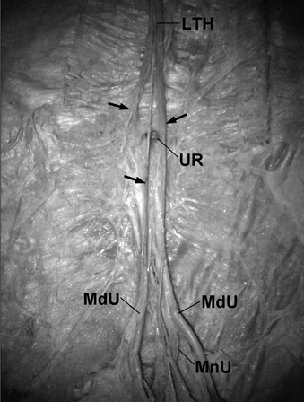
Fig. 2 Photograph showed an umbilical ring (UR) of Type A and umbilical ligaments of Type II. The ligamentum teres hepatis (LTH) divided into 3 branches (arrows), 1 of which attaches lateral to the oval UR and another that crosses the UR and joins the medial umbilical ligament (MdU). The median umbilical ligament (MnU) joins the MdU that is on the right side.

Fig. 3 Photograph of a backlit section showing an umbilical ring (UR) of Type B and umbilical ligaments of Type II. No light is transmitted in the area of the UR because the ring is obliterated. The median umbilical ligament (MnU) joins the medial umbilical ligament (MdU) that is on the right side, and the ligamentum teres hepatis (LTH) divides into 2 branches. The umbilical fascia (UF) is reflected.
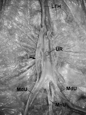
Fig. 4 Photograph showing an umbilical ring (UR) of Type C and umbilical ligaments of Type III. The median umbilical ligament (MnU) and medial umbilical ligament (MdU) fuse into a band that continues to the ligamentum teres hepatis (LTH), covering the UR. A connecting branch (arrow) is evident between the LTH and the MdU that is on the left side.
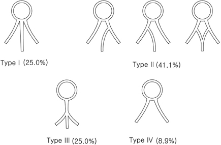
Fig. 5 Four types of umbilical ligaments identified based on their interrelationships.
Reference
-
1. Lemmer JH, Strodel WE, Eckhauser FE. Umbilical hernia incarceration: a complication of medical therapy of ascites. Am J Gastroenterol. 1983. 78:295–296.2. Kives SL, Lara-Torre E. Pyogenic sacroiliitis following an infected umbilical ring. J Pediatr Adolesc Gynecol. 2004. 17:125–129.
Article3. Orda R, Nathan H. Surgical anatomy of the umbilical structures. Int Surg. 1973. 58:458–464.4. Friedrich K, Vogel HM, Henning H. The importance of variant insertions of the ligamentum teres hepatis in the Cruveilhier-Baumgarten syndrome. Endoscopy. 1988. 20:254–259.
Article5. Hammond G, Yglesias L, Davis JE. The urachus, its anatomy and associated fasciae. Anat Rec. 1941. 80:271–287.
Article6. Ikossi DG, Shaheen R, Mallory B. Laparoscopic femoral hernia repair using umbilical ligament as plug. J Laparoendosc Adv Surg Tech A. 2005. 15:197–200.
Article7. Martin BF, Tudor RG. The umbilical and paraumbilical veins of man. J Anat. 1980. 130:305–322.
- Full Text Links
-
- Actions
-
Cited
- CITED
-
- Close
- Share
-
- Similar articles
-
- Abscess Formation Involving the Falciform Ligament and Ligamentum Teres
- Classification of Umbilical Vein Anomalies Based upon Cases in Dead Fetuses
- Minimally Invasive Treatment of Falciform Ligament Abscess in a 25-Day-Old Neonate: A Case Report
- A peculiar liver with surgically and radiologically important variations: a case report
- Umbilical Polyp
