Parenchymal Neurocutaneous Melanosis in Association with Intraventricular Dermoid and Dandy-Walker Variant: A Case Report
- Affiliations
-
- 1Department of Radiology, The Catholic University of Korea, College of Medicine, Gyeonggi-do, Korea. violet2@catholic.ac.kr
- 2Department of Pathology, The Catholic University of Korea, College of Medicine, Gyeonggi-do, Korea.
- 3Department of Neurosurgery, The Catholic University of Korea, College of Medicine, Gyeonggi-do, Korea.
- KMID: 1782188
- DOI: http://doi.org/10.3348/kjr.2006.7.2.145
Abstract
- Neurocutaneous melanosis (NCM) is a rare congenital disease that is characterized by the presence of large or multiple congenital melanocytic nevi and melanotic lesions of the central nervous system. We report here on the CT and MR imaging findings of an unusual case of NCM that was associated with intraventricular dermoid and Dandy-Walker malformation.
Keyword
MeSH Terms
-
Tomography, X-Ray Computed
Neurocutaneous Syndromes/*epidemiology/radiography
Melanosis/*epidemiology/radiography
Male
Magnetic Resonance Imaging
Humans
Dermoid Cyst/*epidemiology/radiography
Dandy-Walker Syndrome/*epidemiology
Comorbidity
Cerebral Ventricle Neoplasms/*epidemiology/radiography
Arachnoid Cysts/epidemiology/pathology
Adult
Figure
-
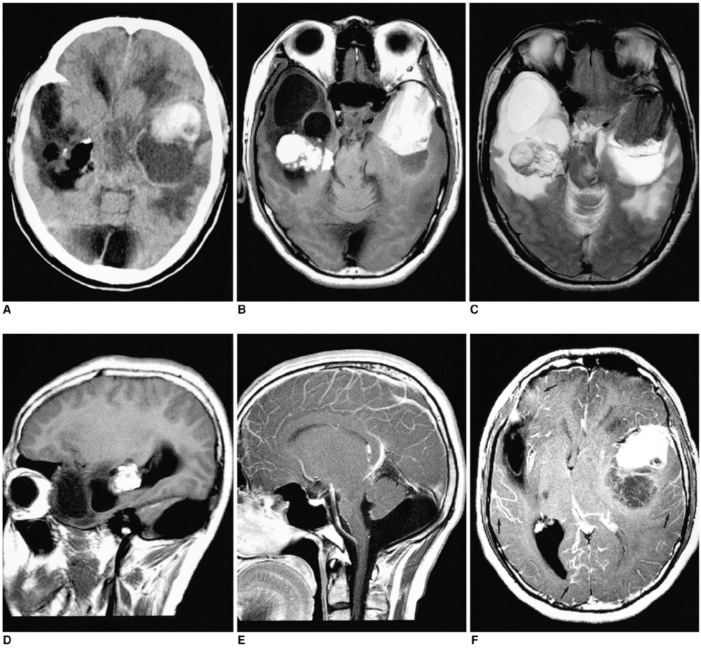
Fig. 1 Neurocutaneous melanosis in a 27-year-old man. A. Noncontrast CT scan demonstrates a hyperdense mass with an adjacent cyst in the left temporal lobe. The CT scan also shows an irregular fatty mass (-105 HU) with marginal calcifications within the temporal horn of the right lateral ventricle. B, C. The axial T1-weighted (B) and T2-weighted (C) MR images show a left temporal lobe mass that is hyperintense on T1-weighted images and it is hypointense on T2-weighted images. There is a peritumoral cyst posterior to the main mass. The MR images also showed a mass in the right lateral ventricle, which appears homogeneously hyperintense on the T1-weighted images and heterogeneously hyperintense on the T2-weighted images; this is consistent with a dermoid cyst. The cystic encephalomalacia in the right temporal lobe is probably related to an early childhood insult. D. The right parasagittal T1-weighted MR image confirms the location of the right side mass within the temporal horn of the lateral ventricle. E. The midline sagittal contrast-enhanced T1-weighted MR image reveals hypoplasia of the inferior vermis and dilatation of the inferior fourth ventricle that communicates to the enlarged posterior fossa. F. The axial contrast-enhanced T1-weighted MR image shows mild enhancements of the wall of the peritumoral cyst in the left temporal lobe. Also noted is mild diffuse enhancement of the leptomeninges (arrows).
Reference
-
1. Fox H. Vinken PJ, Bruyn GW, editors. Neurocutaneous melanosis. Handbook of clinical neurology. 1972. Amsterdam: North Holland;414–428.2. Byrd SE, Darling CF, Tomita T, Chou P, de Leon GA, Radkowski MA. MR imaging of symptomatic neurocutaneous melanosis in children. Pediatr Radiol. 1997. 27:39–44.3. Barkovich AJ, Frieden IJ, Williams ML. MR of neurocutaneous melanosis. AJNR Am J Neuroradiol. 1994. 15:859–867.4. Demirci A, Kawamura Y, Sze G, Duncan C. MR of parenchymal neurocutaneous melanosis. AJNR Am J Neuroradiol. 1995. 16:603–606.5. Kadonaga JN, Frieden IJ. Neurocutaneous melanosis: definition and review of the literature. J Am Acad Dermatol. 1991. 24:747–755.6. Gomori JM, Grossman RI, Shields JA, Augsburger JJ, Joseph PM, DeSimeone D. Choroidal melanomas: correlation of NMR spectroscopy and MR imaging. Radiology. 1986. 158:443–445.7. Ogawa R, Aoki R, Hyakusoku H. A rare case of intracranial metastatic amelanotic melanoma with cyst. J Clin Pathol. 2003. 56:548–551.8. Kadonaga JN, Barkovich AJ, Edwards MS, Frieden IJ. Neurocutaneous melanosis in association with the Dandy-Walker complex. Pediatr Dermatol. 1992. 9:37–43.9. Kasantikul V, Shuangshoti S, Pattanaruenglai A, Kaoroptham S. Intraspinal melanotic arachnoid cyst and lipoma in neurocutaneous melanosis. Surg Neurol. 1989. 31:138–141.10. Smirniotopoulos JG, Chiechi MV. Teratomas, dermoids, and epidermoids of the head and neck. Radiographics. 1995. 15:1437–1455.
- Full Text Links
-
- Actions
-
Cited
- CITED
-
- Close
- Share
-
- Similar articles
-
- Dandy-Walker Malformation Associated with Neurocutaneous Melanosis
- A Case of Neurocutaneous Giant Melanosis Associated with Dandy-Walker Syndrome
- Neurocutaneous Melanosis in Association with Dandy-Walker Complex with Extensive Intracerebral and Spinal Cord Involvement
- A Case of Congenital Onychodysplasia of the Index Fingers Associated with Dandy-Walker Variant
- A Case of Symptomatic Neurocutaneous Melanosis Associated with the Dandy Walker Complex and Excellent Response of Seizures to Topiramate
