Evaluation of Morphological Plasticity in the Cerebella of Basketball Players with MRI
- Affiliations
-
- 1Department of Anatomy, College of Medicine, Korea University, Korea. irhyu@korea.ac.kr
- 2Department of Exercise Physiology, Graduate School, Korea.
- 3Institute of Sports Science, Korea University, Korea.
- 4Department of Diagnostic Radiology, College of Medicine, Korea University, Korea.
- 5Department of Anatomy, College of Medicine, Yonsei University, Seoul, Korea.
- KMID: 1781847
- DOI: http://doi.org/10.3346/jkms.2006.21.2.342
Abstract
- Cerebellum is a key structure involved in motor learning and coordination. In animal models, motor skill learning increased the volume of molecular layer and the number of synapses on Purkinje cells in the cerebellar cortex. The aim of this study is to investigate whether the analogous change of cerebellar volume occurs in human population who learn specialized motor skills and practice them intensively for a long time. Magnetic resonance image (MRI)-based cerebellar volumetry was performed in basketball players and matched controls with V-works image software. Total brain volume, absolute and relative cerebellar volumes were compared between two groups. There was no significant group difference in the total brain volume, the absolute and the relative cerebellar volume. Thus we could not detect structural change in the cerebellum of this athlete group in the macroscopic level.
MeSH Terms
Figure
-
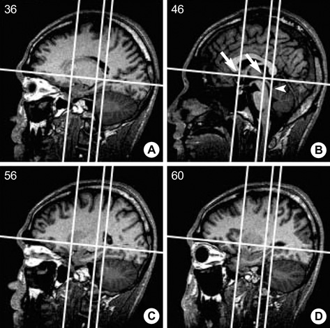
Fig. 1 Landmark-based separation of the cerebellar peduncles from the cerebellar white matter and the brainstem. On the mid-sagittal slice (slice 46/90 in this subject), perpendicular lines are drawn at a 90 degree angle to the bi-commissural line at the AC and PC (arrows) and through the posterior border of the inferior colliculus (arrowhead). These lines are superimposed on all sagittal slices and the latter perpendicular line is used to differentiate the cerebellar peduncles from the brainstem. (slice numbers are placed on top left corner of each slice).
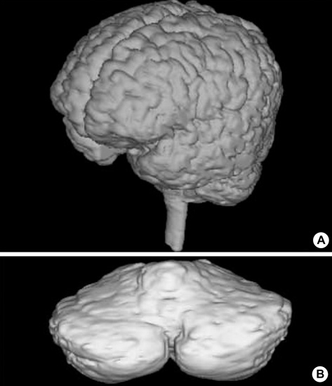
Fig. 2 3-D model of the total brain (A) and the absolute cerebellum (B) using 3-D medical software package for tBV, aCV, rCV.
Cited by 1 articles
-
Is There Relationship between Brain Atrophy and Higher Incidence of Hip Fracture in Old Age? -A Preliminary Study-
Tae Hoon Kim, Seung Woo Suh, Jin-Ho Hwang, Tae Hwan Yoon
Yonsei Med J. 2013;54(6):1511-1515. doi: 10.3349/ymj.2013.54.6.1511.
Reference
-
1. Black JE, Isaacs KR, Anderson BJ, Alcantara AA, Greenough WT. Learning causes synaptogenesis, whereas motor activity causes angiogenesis, in cerebellar cortex of adult rats. Proc Natl Acad Sci USA. 1990. 87:5568–5572.
Article2. Kleim JA, Swain RA, Czerlanis CM, Kelly JL, Pipitone MA, Greenough WT. Learning dependent dendritic hypertrophy of cerebellar stellate cells: plasticity of local circuit neurons. Neurobiol Learn Mem. 1997. 67:29–33.3. Mauk MD, Garcia KS, Medina JF, Steele PM. Does cerebellar LTD mediate motor learning? Toward a resolution without a smoking gun. Neuron. 1998. 20:359–362.
Article4. Thach WT. A role for the cerebellum in learning movement coordination. Neurobiol Learn Mem. 1998. 70:177–188.
Article5. Kim SG, Ugurbil K, Strick PL. Activation of a cerebellar output nucleus during cognitive processing. Science. 1994. 265:949–951.
Article6. Raichle ME, Fiez JA, Videen TO, MacLeod AM, Pardo JV, Fox PT, Petersen SE. Practice-related changes in human brain functional anatomy during nonmotor learning. Cereb Cortex. 1994. 4:8–26.
Article7. Fiez JA, Raife EA, Balota DA, Schwarz JP, Raichle ME, Petersen SE. A positron emisson tomography study of the short-term maintenance of verbal information. J Neurosci. 1996. 16:808–822.8. Gao JH, Parsons LM, Bower JM, Xiong J, Li J, Fox PT. Cerebellum implicated in sensory acquisition and discrimination rather than motor control. Science. 1996. 272:545–547.
Article9. Allen G, Buxton RB, Wong EC, Courchesne E. Attentional activation of the cerebellum independent of motor involvement. Science. 1997. 725:1940–1942.
Article10. Kleim JA, Swain RA, Armstrong KA, Napper RM, Jones TA, Greenough WT. Selective synaptic plasticity within the cerebellar cortex following complex motor skill learning. Neurobiol Learn Mem. 1998. 69:274–289.
Article11. Pysh JJ, Weiss GM. Exercise during development induces an increase in Purkinje cell dendritic tree size. Science. 1979. 206:230–232.
Article12. Paradiso S, Andreaen NC, O'Leary DS, Arndt S, Robinson RG. Cerebellar size and cognition: correlations with IQ, verbal memory and motor dexterity. Neuropsychiatry Neuropsychol Behav Neurol. 1997. 10:1–8.13. Reiss AL, Abrams MT, Singer HS, Ross JL, Denckla MB. Brain development, gender and IQ in children-a volumetric imaging study. Brain. 1996. 119:1763–1774.14. Rhyu IJ, Cho TH, Lee NJ, Uhm CS, Kim H, Suh YS. Magnetic resonance image-based cerebellar volumetry in healthy Korean adults. Neurosci Lett. 1999. 270:149–152.
Article15. Chung SC, Choi DY, Lee BY, Lee BS, Eom JS, Sohn JH. Measuring of the cerebellar volume of normal Koreans in their 20s and 40s using magnetic resonance imaging. J Korean Radiol Soc. 2004. 51:489–493.
Article16. Chung SC, Lee BY, Tack GR, Lee SY, Eom JS, Sohn JH. Effects of age, gender, and weight on cerebellar volume of Korean people. Brain Res. 2005. 1042:233–235.17. Schlaug G, Lee LH, Thangraj V, Edelman RR, Warach S. Macrostructural adapation of the cerebellum in musicians. Soc Neurosci Abstr. 1998. 24:2118. (842.7).18. Hutchinson S, Lee LH, Gaab N, Schlaug G. Cerebellar volume of musicians. Cereb Cortex. 2003. 13:943–949.
Article19. Kim HJ, Yoon HR, Kim KD, Kang MK, Kwak HH, Park HD, Han SH, Park CS. Personal-Computer-based three dimensional reconstruction and simulation of maxillary sinus. Surg Radiol Anat. 2003. 24:393–399.20. Huang Y, Knorr U, Schlaug G, Seitz RJ, Steinmetz H. Segmentation of MR images for partial-volume-effect correction and individual integration with PET images of the human brain. J Cereb Blood Flow Metab. 1993. 13:S315.21. Peters M, Jancke L, Zilles K. Comparison of overall brain volume and midsagittal corpus callosum surface area as obtained from NMR scans and direct anatomical measures: a within-subject study on autopsy brains. Neuropsychologia. 2000. 38:1375–1381.
Article22. Courchesne E, Press GA, Murakami J, Berthoty D, Grafe M, Wiley CA, Hesselink JR. The cerebellum in sagittal plane-anatomic-MR correlation: 1. The vermis. AJR Am J Roentgehol. 1989. 153:829–835.
Article23. Press GA, Murakami J, Courchesne E, Berthoty DP, Grafe M, Wiley CA, Hesselink JR. The cerebellum in sagittal plane anatomic MR correlation: 2. The cerebellar hemispheres. AJR Am J Roentgenol. 1989. 153:837–846.24. Filipek PA, Richelme C, Kennedy DN, Caviness VS Jr. The young adult human brain: an MRI-based morphometric analysis. Cereb Cortex. 1994. 4:344–360.
Article25. Luft AR, Skalej M, Welte D, Kolb R, Burk K, Schulz JB, Klockgether T, Voigt K. A new semiautomated, three-dimensional technique allowing precise quantification of total and regional cerebellar volume using MRI. Magn Reson Med. 1998. 40:143–151.
Article26. Witelson SF. Hand and sex differences in the isthmus and genu of the human corpus callosum: a post-mortem morphological study. Brain. 1989. 112:799–835.27. Peters M. Sex differences in human brain size and the general meaning of differences in brain size. Can J Psychol. 1991. 45:507–522.
Article28. Witelson SF, Kigar DL, Harvey T. The exceptional brain of Albert Einstein. Lancet. 1999. 353:2149–2153.
Article29. Woodruff-Pak DS, Vogel RW III, Ewers M, Coffey J, Boyko OB, Lemieux SK. MRI-assessed volume of cerebellum correlates with associative learning. Neurobiol Learn Mem. 2001. 76:342–357.
Article30. Praag VH, Kempermann G, Gage FH. Running increases cell proliferation and neurogenesis in the adult mouse dentate gyrus. Nat Neurosci. 1999. 2:266–270.
Article
- Full Text Links
-
- Actions
-
Cited
- CITED
-
- Close
- Share
-
- Similar articles
-
- Cerebral Cortex Changes in Basketball Players
- The Comparison of Psychological Characteristics between Korean and Japanese Women Pro-Basketball Players
- Comparison of Temperament and Cognitive Function Between Basketball and Baseball Players
- Evaluation of Pain and Ultrasonography on Shoulder in Poliomyelitis Wheelchair Basketball Players
- Comparison of Isokinetic Knee Strength Profiles According to History of Knee Surgery in Korean Women’s Professional Basketball Players
