Acute Geometric Changes of the Mitral Annulus after Coronary Occlusion: A Real-Time 3D Echocardiographic Study
- Affiliations
-
- 1Department of Cardiology, Inha University Hospital, Inchon, Korea. kuonmd@inha.ac.kr
- 2Department of Pathology, Korea University College of Medicine, Seoul, Korea.
- 3Cardiovascular Imaging Center, Department of Cardiology, The Cleveland Clinic Foundation, Cleveland, Ohio, U.S.A.
- 4National Heart, Lung and Blood Institute, Bethesda, Maryland, U.S.A.
- KMID: 1781825
- DOI: http://doi.org/10.3346/jkms.2006.21.2.217
Abstract
- We performed real-time 3D echocardiography in sixteen sheep to compare acute geometric changes in the mitral annulus after left anterior descending coronary artery (LAD, n=8) ligation and those after left circumflex coronary artery (LCX, n=8) ligation. The mitral regurgitation (MR) was quantified by regurgitant volume (RV) using the proximal isovelocity surface area method. The mitral annulus was reconstructed through the hinge points of the annulus traced on 9 rotational apical planes (angle increment=20 degrees). Mitral annular area (MAA) and the ratio of antero-posterior (AP) to commissure-commissure (CC) dimension of the annulus were calculated. Non-planar angle (NPA) representing non-planarity of the annulus was measured. After LCX occlusion, there were significant increases of the MAA during both early and late systole (p<0.01) with significant MR (RV: 30+/-14 mL), while there was neither a significant increase of MAA, nor a significant MR (RV: 4+/-5 mL) after LAD occlusion. AP/CC ratio (p<0.01) and NPA (p<0.01) also significantly increased after LCX occlusion during both early and late systole. The mitral annulus was significantly enlarged in the antero-posterior direction with significant decrease of non-planarity compared to LAD occlusion immediately after LCX occlusion.
MeSH Terms
Figure
-
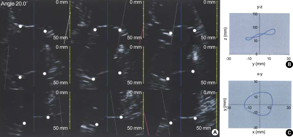
Fig. 1 Nine rotational apical planes were used to trace 18 hinge points (white dots) of the mitral annulus (A). 3D shape of the mitral annulus reconstructed from the 18 hinge points (B). Projected view of 3D reconstructed annulus (C).
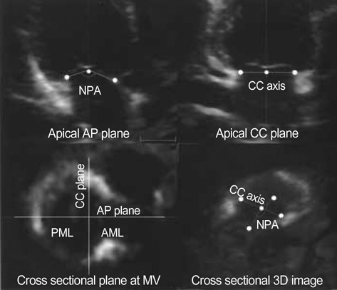
Fig. 2 Volumetric image showing how the non-planar angle (NPA) of the annulus was measured. Using 3D computer software (TomTec), we measured the angle between two vectors from two hinge points of the annulus (white dots) in the antero-posterior (AP) plane to the center of the axis connecting two commssures (white dot) in the commissure-commissure (CC) plane. AML, anterior mitral leaflet; MV, mitral valve; PML, posterior mitral leaflet.

Fig. 3 Change of the non-planar angle (NPA) of the mitral annulus after coronary occlusion.

Fig. 4 Graphs showing significant correlations (p<0.01) of percent change (Δ) of the non-planar angle (NPA) with that of the mitral annular area (MAA) and the antero-posterior dimension (AP).

Fig. 5 Graphs showing weak but statistically insignificant correlations (p>0.05) of percent change (Δ) of the mitral annular area (MAA) and the non-planar angle (NPA) with regurgitant volume (RV).

Fig. 6 Apical movement of the posterior annulus (arrow) before and after LCX occlusion. AP, antero-posterior dimension; NPA, non-planar angle. Note the smaller movement of the posterior annulus after LCX occlusion than before the occlusion.
Reference
-
1. Gahl K, Sutton R, Pearson M, Caspari P, Lairet A, McDonald L. Mitral regurgitation in coronary heart disease. Br Heart J. 1977. 39:13–18.
Article2. Hendren WG, Nemec JJ, Lytle BW, Loop FD, Taylor PC, Stewart RW, Cosgrove DM 3rd. Mitral valve repair for ischemic mitral insufficiency. Ann Thorac Surg. 1991. 52:1246–1251.
Article3. Czer LS, Maurer G, Trento A, DeRobertis M, Nessim S, Blanche C, Kass RM, Chaux A, Matloff JM. Comparative efficacy of ring and suture annuloplasty for ischemic mitral regurgitation. Circulation. 1992. 86:II46–II52.4. Dion R. Ischemic mitral regurgitation: When and how should it be corrected? J Heart Valve Dis. 1993. 2:536–543.5. Timek T, Glasson JR, Dagum P, Green GR, Nistal JF, Komeda M, Daughters GT, Bolger AF, Foppiano LE, Ingels NB Jr, Miller DC. Ring annuloplasty prevents delayed leaflet coaptation and mitral regurgitation during acute left ventricular ischemia. J Thorac Cardiovasc Surg. 2000. 119:774–783.
Article6. Glasson JR, Komeda M, Daughters GT, Bolger AF, Karlsson MO, Foppiano LE, Hayase M, Oesterle SN, Ingels NB Jr, Miller DC. Early systolic mitral leaflet "loitering" during acute ischemic mitral regurgitation. J Thorac Cardiovasc Surg. 1998. 116:193–205.
Article7. Gorman JH 3rd, Gorman RC, Jackson BM, Hiramatsu Y, Gikakis N, Kelley ST, Sutton MG, Plappert T, Edmunds LH Jr. Distortions of the mitral valve in acute ischemic mitral regurgitation. Ann Thorac Surg. 1997. 64:1026–1031.
Article8. Levine RA, Handschumacher MD, Sanfilippo AJ, Hagege AA, Harrigan P, Marshall JE, Weyman AE. Three-dimensional echocardiographic reconstruction of the mitral valve, with implications for the diagnosis of mitral valve prolapse. Circulation. 1989. 80:589–598.
Article9. Salustri A, Becker AE, van Herwerden L, Vletter WB, Ten Cate FJ, Roelandt JR. Three-dimensional echocardiography of the normal and pathologic mitral valve: a comparison with two-dimensional transesophageal echocardiography. J Am Coll Cardiol. 1996. 27:1502–1510.10. Glasson JR, Komeda M, Daughters GT 2nd, Bolger AF, MacIsaac A, Oesterle SN, Ingels NB Jr, Miller DC. Three-dimensional dynamics of the canine mitral annulus during ischemic mitral regurgitation. Ann Thorac Surg. 1996. 62:1059–1067.
Article11. Komeda M, Glasson JR, Bolger AF, Daughters GT 2nd, MacIsaac A, Oesterle SN, Ingels NB Jr, Miller DC. Geometric determinants of ischemic mitral regurgitation. Circulation. 1997. 96:128–133.12. Dagum P, Timek TA, Green GR, Lai D, Daughters GT, Liang DH, Hayase M, Ingels NB Jr, Miller DC. Coordinate-free analysis of mitral valve dynamics in normal and ischemic hearts. Circulation. 2000. 102:III62–III69.
Article13. Otsuji Y, Handschumacher MD, Liel-Cohen N, Tanabe H, Jiang L, Schwammenthal E, Guerrero JL, Nicholls LA, Vlahakes GJ, Levine RA. Mechanism of ischemic mitral regurgitation with segmental left ventricular dysfunction: three-dimensional echocardiographic studies in models of acute and chronic progressive regurgitation. J Am Coll Cardiol. 2001. 37:641–648.
Article14. Binder TM, Rosenhek R, Porenta G, Maurer G, Baumgartner H. Improved assessment of mitral valve stenosis by volumetric real-time three-dimensional echocardiography. J Am Coll Cardiol. 2000. 36:1355–1361.
Article15. Shiota T, Jones M, Chikada M, Fleishman CE, Castellucci JB, Cotter B, DeMaria AN, von Ramm OT, Kisslo J, Ryan T, Sahn DJ. Real-time three-dimensional echocardiography for determining right ventricular stroke volume in an animal model of chronic right ventricular volume overload. Circulation. 1998. 97:1897–1900.
Article16. Schiller NB, Shah PM, Crawford M, DeMaria A, Devereux R, Feigenbaum H, Gutgesell H, Reichek N, Sahn D, Schnittger I, Silverman NH, Tajik AJ. Recommendations for quantitation of the left ventricle by two-dimensional echocardiography. American Society of Echocardiography Committee on Standards, Subcommittee on Quantitation of Two-dimensional Echocardiograms. J Am Soc Echocardiogr. 1989. 2:358–367.17. Aotsuka H, Tobita K, Hamada H, Uchishiba M, Tateno S, Matsuo K, Fujiwara T, Niwa K. Validation of the proximal isovelocity surface area method for assessing mitral regurgitation in children. Pediatr Cardiol. 1996. 17:351–359.
Article18. Flachskampf FA, Chandra S, Gaddipatti A, Levine RA, Weyman AE, Ameling W, Hanrath P, Thomas JD. Analysis of shape and motion of the mitral annulus in subjects with and without cardiomyopathy by echocardiographic 3-dimensional reconstruction. J Am Soc Echocardiogr. 2000. 13:277–287.
Article19. Pai RG, Tanimoto M, Jintapakorn W, Azevedo J, Pandian NG, Shah PM. Volume-rendered three-dimensional dynamic anatomy of the mitral annulus using a transesophageal echocardiographic technique. J Heart Valve Dis. 1995. 4:623–627.20. Gorman RC, McCaughan JS, Ratcliffe MB, Gupta KB, Streicher JT, Ferrari VA, St John-Sutton MG, Bogen DK, Edmunds LH Jr. Pathogenesis of acute ischemic mitral regurgitation in three dimensions. J Thorac Cardiovasc Surg. 1995. 109:684–693.
Article21. Boltwood CM, Tei C, Wong M, Shah PM. Quantitative echocardiography of the mitral complex in dilated cardiomyopathy: the mechanism of functional mitral regurgitation. Circulation. 1983. 68:498–508.
Article22. Kono T, Sabbah HN, Rosman H, Alam M, Jafri S, Stein PD, Goldstein S. Mechanism of functional mitral regurgitation during acute myocardial ischemia. J Am Coll Cardiol. 1992. 19:1101–1105.
Article23. Sabbah HN, Kono T, Rosman H, Jafri S, Stein PD, Goldstein S. Left ventricular shape: a factor in the etiology of functional mitral regurgitation in heart failure. Am Heart J. 1992. 123:961–966.
Article24. Ormiston JA, Shah PM, Tei C, Wong M. Size and motion of the mitral valve annulus in man. I. A two-dimensional echocardiographic method and findings in normal subjects. Circulation. 1981. 64:113–120.
Article25. He S, Lemmon JD Jr, Weston MW, Jensen MO, Levine RA, Yoganathan AP. Mitral valve compensation for annular dilatation: in vitro study into the mechanisms of functional mitral regurgitation with an adjustable annulus model. J Heart Valve Dis. 1999. 8:294–302.26. Yiu SF, Enriquez-Sarano M, Tribouilloy C, Seward JB, Tajik AJ. Determinants of the degree of functional mitral regurgitation in patients with systolic left ventricular dysfunction: A quantitative clinical study. Circulation. 2000. 102:1400–1406.27. Kwan J, Shiota T, Agler DA, Popovic ZB, Qin JX, Gillinov MA, Stewart WJ, Cosgrove DM, McCarthy PM, Thomas JD. Geometric differences of the mitral apparatus between ischemic and dilated cardiomyopathy with significant mitral regurgitation: real-time three-dimensional echocardiography study. Circulation. 2003. 107:1135–1140.28. Kaplan SR, Bashein G, Sheehan FH, Legget ME, Munt B, Li XN, Sivarajan M, Bolson EL, Zeppa M, Arch MZ, Martin RW. Three-dimensional echocardiographic assessment of annular shape changes in the normal and regurgitant mitral valve. Am Heart J. 2000. 139:378–387.
Article29. Kwan J, Qin JX, Popovic ZB, Agler DA, Thomas JD, Shiota T. Geometric changes of mitral annulus assessed by real-time 3-dimensional echocardiography: becoming enlarged and less nonplanar in the anteroposterior direction during systole in proportion to global left ventricular systolic function. J Am Soc Echocardiogr. 2004. 17:1179–1184.
Article30. Shiota T, Jones M, Teien DE, Yamada I, Passafini A, Ge S, Sahn DJ. Dynamic change in mitral regurgitant orifice area: comparison of color Doppler echocardiographic and electromagnetic flowmeter-based methods in a chronic animal model. J Am Coll Cardiol. 1995. 26:528–536.
Article31. Shiota T, Jones M, Teien DE, Yamada I, Passafini A, Ge S, Shandas R, Valdes-Cruz LM, Sahn DJ. Evaluation of mitral regurgitation using a digitally determined color Doppler flow convergence 'centerline' acceleration method. Studies in an animal model with quantified mitral regurgitation. Circulation. 1994. 89:2879–2887.
Article
- Full Text Links
-
- Actions
-
Cited
- CITED
-
- Close
- Share
-
- Similar articles
-
- Does The Mitral Annulus Shrink or Enlarge During Systole? A Real-Time 3D Echocardiography Study
- Role of modern 3D echocardiography in valvular heart disease
- Assessment of Ventricular Function Using Tissue Doppler Imaging in Kawasaki Disease
- Effects of Decreased Annular Height and Annular Saddle-Shaped Non-Planarity in Degenerative Severe Mitral Regurgitation with Normal Left Ventricular Ejection Fraction: Real-Time 3D Transesophageal Echocardiography
- Assessment of Diastolic Function Using Mitral Annulus Velocity by Doppler Tissue Velocity in the Patients with Left Ventricular Hypertrophy
