Does The Mitral Annulus Shrink or Enlarge During Systole? A Real-Time 3D Echocardiography Study
- Affiliations
-
- 1Department of Cardiology, Inha University Hospital, Incheon, Korea. kuonmd@inha.ac.kr
- KMID: 1779119
- DOI: http://doi.org/10.3346/jkms.2009.24.2.203
Abstract
- This study was conducted to explore the geometrical changes of the mitral annulus during systole. The 3D shape of the mitral annulus was reconstructed in 13 normal subjects who had normal structure of the mitral apparatus using real-time 3D echocardiography (RT3DE) and 3D computer software. The two orthogonal (antero-posterior and commissure-commissure) dimensions, the areas (2D projected and 3D surface) and the non-planarity of the mitral annulus were estimated during early, mid and late systole. We demonstrated that the MA had a "saddle shape" appearance and it consistently enlarged mainly in the antero-posterior direction from early to late systole with lessening of its non-planarity, as was determined by 3D reconstruction using RT3DE and 3D computer software.
Keyword
MeSH Terms
Figure
-

Fig. 1 Generation of 16 rotational apical planes and tracing 32 hinge points of the mitral annulus (white spots) on each plane for 3D reconstruction of the annulus using newly developed 3D computer software.
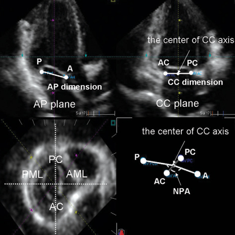
Fig. 2 Definition of anatomical reference markers of the mitral annulus on the cross sectional volumetric plane and geometrical measurements of the annulus. A, anterior point; P, posterior point; AC, anterior commissure; PC, posterior commissure; AML, anterior mitral leaflet; PML, posterior mitral leaflet; AP, antero-posterior; CC, commissure-commissure; NPA, non-planar angle.

Fig. 3 Division of the mitral annulus into anterior and posterior annuli by the CC axis and the NPA measurement. CC, commissure-commissure; NPA, non-planar angle.
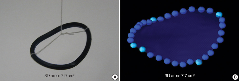
Fig. 4 3D reconstruction and 3D area calculation (B) of a "saddle-shaped" artificial structure (A) using newly developed 3D computer software for its validation.

Fig. 5 Anterior oblique (left) and projected (right) views of the mitral annulus. A, anterior point; P, posterior point; AC, anterior commissure; PC, posterior commissure.
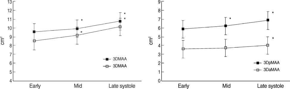
Fig. 6 Line graphs showing changes of the areas (2D and 3D) of the mitral annulus during three phases (early, mid, and late) of the systole. 2DMAA, 2D area of the annulus; 3DMAA, 3D area of the annulus; 3DaMAA, 3D area of the anterior annulus; 3DpMAA, 3D area of the posterior annulus. *p<0.05 versus prior phase by post-hoc comparison test (Bonferroni's method).

Fig. 7 Change of the AP dimension of the mitral annulus from early to late systole in comparison with that of the AP diameter of the left ventricle. AP, antero-posterior.

Fig. 8 Change of the 3D shape of the mitral annulus from early to late systole. NPA, non-planar angle.
Reference
-
1. Tsakiris AG, Von Bernuth G, Rastelli GC, Bourgeois MJ, Titus JL, Wood EH. Size and motion of the mitral valve annulus in anesthetized intact dogs. J Appl Physiol. 1971. 30:611–618.
Article2. Ormiston JA, Shah PM, Tei C, Wong M. Size and motion of the mitral valve annulus in man. I. A two-dimensional echocardiographic method and findings in normal subjects. Circulation. 1981. 64:113–120.
Article3. Komoda T, Hetzer R, Uyama C, Siniawski H, Maeta H, Rosendahl UP, Ozaki K. Mitral annular function assessed by 3D imaging for mitral valve surgery. J Heart Valve Dis. 1994. 3:483–490.4. Pai RG, Tanimoto M, Jintapakorn W, Azevedo J, Pandian NG, Shah PM. Volume-rendered three-dimensional dynamic anatomy of the mitral annulus using a transesophageal echocardiographic technique. J Heart Valve Dis. 1995. 4:623–627.5. Glasson JR, Komeda M, Daughters GT, Niczyporuk MA, Bolger AF, Ingels NB, Miller DC. Three-dimensional regional dynamics of the normal mitral anulus during left ventricular ejection. J Thorac Cardiovasc Surg. 1996. 111:574–585.
Article6. Flachskampf FA, Chandra S, Gaddipatti A, Levine RA, Weyman AE, Ameling W, Hanrath P, Thomas JD. Analysis of shape and motion of the mitral annulus in subjects with and without cardiomyopathy by echocardiographic 3-dimensional reconstruction. J Am Soc Echocardiogr. 2000. 13:277–287.
Article7. Nilsson B, Bojo L, Wandt B. Influence of body size and age on maximal systolic velocity of mitral annulus motion. Clin Physiol. 2000. 20:272–278.
Article8. Emilsson K, Alam M, Wandt B. The relation between mitral annulus motion and ejection fraction: a nonlinear function. J Am Soc Echocardiogr. 2000. 13:896–901.
Article9. Kwan J, Qin JX, Popovic ZB, Agler DA, Thomas JD, Shiota T. Geometric changes of mitral annulus assessed by real-time 3-dimensional echocardiography: becoming enlarged and less nonplanar in the anteroposterior direction during systole in proportion to global left ventricular systolic function. J Am Soc Echocardiogr. 2004. 17:1179–1184.
Article10. Qin JX, Shiota T, Tsujino H, Saracino G, White RD, Greenberg NL, Kwan J, Popovic ZB, Agler DA, Stewart WJ, Thomas JD. Mitral annular motion as a surrogate for left ventricular ejection fraction: real-time three-dimensional echocardiography and magnetic resonance imaging studies. Eur J Echocardiogr. 2004. 5:407–415.
Article11. Ahmad RM, Gillinov AM, McCarthy PM, Blackstone EH, Apperson-Hansen C, Qin JX, Agler D, Shiota T, Cosgrove DM. Annular geometry and motion in human ischemic mitral regurgitation: novel assessment with three-dimensional echocardiography and computer reconstruction. Ann Thorac Surg. 2004. 78:2063–2068.
Article12. Watanabe N, Ogasawara Y, Yamaura Y, Kawamoto T, Akasaka T, Yoshida K. Geometric deformity of the mitral annulus in patients with ischemic mitral regurgitation: a real-time three-dimensional echocardiographic study. J Heart Valve Dis. 2005. 14:447–452.13. Watanabe N, Ogasawara Y, Yamaura Y, Wada N, Kawamoto T, Toyota E, Akasaka T, Yoshida K. Mitral annulus flattens in ischemic mitral regurgitation: geometric differences between inferior and anterior myocardial infarction: a real-time 3-dimensional echocardiographic study. Circulation. 2005. 112:9 Suppl. I458–I462.14. Timek TA, Lai DT, Dagum P, Green GR, Glasson JR, Daughters GT, Ingels NB Jr, Miller DC. Mitral annular dynamics during rapid atrial pacing. Surgery. 2000. 128:361–367.
Article15. Davis PK, Kinmonth JB. The movements of the annulus of the mitral valve. J Cardiovasc Surg (Torino). 1963. 4:427–431.
- Full Text Links
-
- Actions
-
Cited
- CITED
-
- Close
- Share
-
- Similar articles
-
- Acute Geometric Changes of the Mitral Annulus after Coronary Occlusion: A Real-Time 3D Echocardiographic Study
- Advances in the Evaluation of Mitral Complex Geometry:Insights from Transthoracic Real-time Three-dimensional Echocardiography
- Role of modern 3D echocardiography in valvular heart disease
- Echocardiographic Assessment of Mitral Valve Regurgitation
- Effects of Decreased Annular Height and Annular Saddle-Shaped Non-Planarity in Degenerative Severe Mitral Regurgitation with Normal Left Ventricular Ejection Fraction: Real-Time 3D Transesophageal Echocardiography
