Lipogranuloma with Osseous Metaplasia in the Breast That Developed after "Bu-Hwang" Oriental Medicine Treatment
- Affiliations
-
- 1Department of Diagnostic Radiology, Gachon University Gil Hospital, Incheon, Korea. shpark@gilhospital.com
- 2Department of Surgery, Gachon University Gil Hospital, Incheon, Korea.
- KMID: 1779680
- DOI: http://doi.org/10.3349/ymj.2011.52.2.373
Abstract
- A lipogranuloma is an inflammatory reactive process associated with exogenous or endogenous lipids, and it's occurrence in the breast has rarely been reported. Osseous metaplasia, which is used to describe bone formation in abnormal locations, can develop from several conditions such as trauma or a tumor. However, few studies have reported benign breast lesions that have been seen as osseous metaplasia. We present a case of a benign calcified breast lesion that developed after a traumatic treatment process called "Bu-Hwang", and it was confirmed as a lipogranuloma with osseous metaplasia. To the best of our knowledge, this is the first reported case of a lipogranuloma with osseous metaplasia in the breast.
Keyword
MeSH Terms
Figure
-

Fig. 1 Mammography of the left breast shows an approximate 3.5 cm sized, relatively well-demarcated lesion with a bizarre shape and bright density in the upper medial portion of the left breast. (A) Mediolateral oblique view. (B) Craniocaudal view.
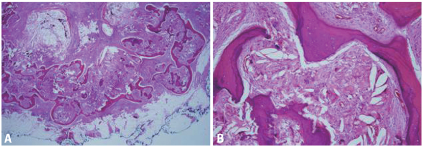
Fig. 2 (A) A photomicrograph shows numerous fat vacuoles with surrounding scattered lymphocytes and foreign body type multinucleated giant cells forming granulomas. (B) Metaplastic bone structures surrounded by fat vacuoles and giant cells are seen [hematoxylin and eosin staining, original magnification ×12.5 (A), ×100 (B)].

Fig. 3 Histopathology shows osteocystes and osteoblast in the bony trabeculae (A), and epithelioid cells in granulomatous changes (B) (heand eosin staining, original magnification ×400).
Reference
-
1. Akbulut M, Utku Y, Soysal S. Lipogranuloma of the cervix in a postmenopausal patient with a uterine prolapse. Arch Gynecol Obstet. 2008. 277:277–279.
Article2. Kayaselçuk F, Kayaselçuk U, Ozerdem OR, Tuncer I. Posttraumatic lipogranuloma of the hand. Ann Plast Surg. 2002. 48:223–224.
Article3. Gal-Gombos EC, Esserman LE, Poniecka AW, Odzer SL, Weisberg S, Godinez J, et al. Osseous metaplasia of the breast: diagnosis with stereotactic core biopsy. Breast J. 2002. 8:50–52.
Article4. Lynch GL, Scagliotti RH. Osseous metaplasia in the eye of a dog. Vet Pathol. 2007. 44:222–224.
Article5. White V, Shaw AG, Tierney GM, Lund JN, Semeraro D. Osseous metaplasia in an ulcerating tubular adenoma of the colon: a case report. J Med Case Reports. 2008. 2:130.
Article6. Sahin A, Tekgül S, Ergen A, Basar I, Dilek H, Ruacan S. Sclerosing lipogranuloma of the penis. A case report. Int Urol Nephrol. 1991. 23:595–598.
Article7. Hopkins KL, Lane B, Zatz LM, Mindelzun RE. Scalp lipogranuloma due to dermal lipid injections: CT and MR findings. AJR Am J Roentgenol. 1995. 165:233.
Article8. Hunter-Craig ID, Tuddenham EG, Earle JH. Lipogranuloma of the breast due to phenothiazine therapy. Br J Surg. 1970. 57:76–79.
Article9. Meyer PN, Al-Masri H, Alkan S. Osseous metaplasia in diffuse large B-cell lymphoma of the kidney. Am J Hematol. 2007. 82:321–324.
Article10. Nishida Y, Kohno N, Furuya Y, Nakatani T, Kaneko S, Sashikata T, et al. [Mammary fibroadenoma showing osseous metaplasia: a case report]. Gan No Rinsho. 1989. 35:1461–1465.
