Radiologic Findings of Renal Hemangioma: Report of Three Cases
- Affiliations
-
- 1Department of Diagnostic Radiology, College of Medicine, Hanyang University, Seoul, Korea.
- KMID: 1777298
- DOI: http://doi.org/10.3348/kjr.2000.1.1.60
Abstract
- Renal hemangioma is an uncommon benign tumor which usually causes painless or painful gross hematuria. Its preoperative diagnosis is extremely difficult or even impossible. We experienced three cases of renal hemangioma, located mainly at the pelvocalyceal junction or in the inner medulla. US demonstrated variable echogenecity, and CT revealed a lack of significant enhancement. Where there is gross hematuria in a young adult, especially when the renal mass located in the pelvocalyceal junction or inner medulla shows little enhancement on CT, renal heman-gioma should form part of the differential diagnosis.
MeSH Terms
Figure
-
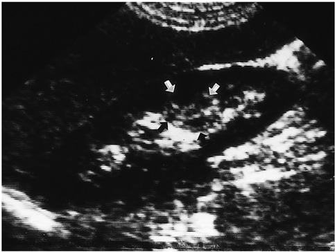
Fig. 1 Renal hemangioma in a 31-years-old man. Longitudinal US shows a well-defined round echogenic mass lesion (arrows) containing multiple anechoic areas in the lower pole of the right kidney.
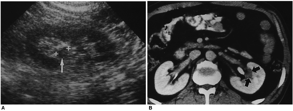
Fig. 2 Renal hemangima in a 31-years-old man. A. US shows a 1.5 cm-sized mass (arrow) in the pelvis of the left kidney, which is isoechoic to renal parenchyma. B. Enhanced CT scan shows a non-enhancing, low attenuated mass with lobulated margin (arrows) adjacent to the pelvis.
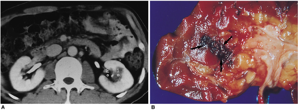
Fig. 3 Renal hemangioma in a 31-years-old man. A. Enhanced CT scan shows a non-enhancing mass with lobulated margin in the lower pole of the left kidney. Unenhanced CT scan shows a high-attenuated mass suggesting hemorrhage (not shown). B. Gross pathologic specimen shows a hemorrhagic lesion (arrows) at the corticomedullary junction of the lower pole.
Cited by 1 articles
-
Hemangioma in Renal Pelvis
Myeong Su Chu
Kosin Med J. 2021;36(2):175-179. doi: 10.7180/kmj.2021.36.2.175.
Reference
-
1. Motley RC, Patterson DE, Weiland LH. Ureteroscopic visualization of a cavernous hemangioma of the renal pelvis. J Urol. 1990; 143:788–790.2. Numan F, Berkmen T, Korman U, et al. Cavernous hemangioma of the kidney: case report. Clin Imaging. 1993; 17:106–108.3. Yazaki T, Takahashi S, Ogawa Y, et al. Large renal hemangioma necessitating nephrectomy. Urology. 1985; 25:302–304.4. Jahn H, Nissen HM. Haemangioma of the urinary tract: review of the literature. Br J Urol. 1991; 68:113–117.5. Wang T, Palazzo JP, Mitchell D, et al. Renal capsular hemangioma. J Urol. 1993; 149:1122–1123.6. Gordon R, Rosenmann E, Barzilay B, et al. Correlation of selective angiography and pathology in cavernous hemangioma of the kidney. J Urol. 1976; 115:608–609.7. Petrson NE, Thompson HT. Renal hemangioma. J Urol. 1971; 105:27–31.8. Stanley RJ, Cubillo E, Mancillajimensez R, et al. Cavernous hemangioma of the kidneys. AJR. 1975; 125:682–687.9. Ekelund L, Gothlin J. Renal hemangiomas. An analysis of 13 cases diagnosed by angiography. AJR. 1975; 125:788–794.10. Yamashita Y, Ueno S, Makita O, et al. Hyperechoic renal tumors: anechoic rim and intratumoral cysts in US differentiation of renal cell carcinoma from angiomyolipoma. Radiology. 1993; 188:179–182.11. Siegel CL, Middleton AD, Teefey SA, et al. Angiomyolipoma and renal cell carcinoma: US differentiation. Radiology. 1996; 198:789–793.12. Mittelstaedt CA. Mittelstaedt CA, editor. Kidney. General Ultrasound. 1992. New York: Churchill Livingstone;p. 947–951.
