Primary Antiphospholipid Antibody Syndrome: Neuro radiologic Findings in 11 Patients
- Affiliations
-
- 1Department of Radiology, Asan Medical Center, University of Ulsan College of Medicine, Seoul, Korea.
- KMID: 1777289
- DOI: http://doi.org/10.3348/kjr.2000.1.1.5
Abstract
OBJECTIVE
To describe the neuroradiologic findings of primary antiphospholipid antibody syndrome (PAPS). MATERIALS AND METHODS: During a recent two-year period, abnormally elevated antiphospholipid antibodies were detected in a total of 751 patients. In any cases in which risk factors for stroke were detected-hypertension, diabetes mellitus, hyperlipidemia, smoking, and the presence of SLE or other connective tissue diseases-PAPS was not diagnosed. Neuroradiologic studies were performed in 11 of 32 patients with PAPS. We retrospectively reviewed brain CT (n = 7), MR (n = 8), and cerebral angiography (n = 8) in 11 patients with special attention to the presence of brain parenchymal lesions and cerebral arterial or venous abnormalities. RESULTS: CT or MR findings of PAPS included nonspecific multiple hyper-inten-sity foci in deep white matter on T2-weighted images (5/11), a large infarct in the territory of the middle cerebral artery (4/11), diffuse cortical atrophy (2/11), focal hemorrhage (2/11), and dural sinus thrombosis (1/11). Angiographic findings were normal (5/8) or reflected either occlusion of a large cerebral artery (2/8) or dural sinus thrombosis (1/8). CONCLUSION: Neuroradiologic findings of PAPS are nonspecific but in young or middle-aged adults who show the above mentioned CT or MR findings, and in whom risk factors for stroke are not present, the condition should be suspected.
MeSH Terms
Figure
-
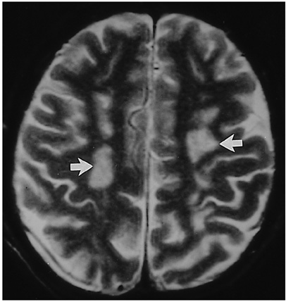
Fig. 1 A 47-year-old man who at the time of examination had suffered headache and general weakness for one month (case 4). Axial T2-weighted MR image shows multiple high signal intensities(arrows) in the white matters at the level of the centrum semiovale.
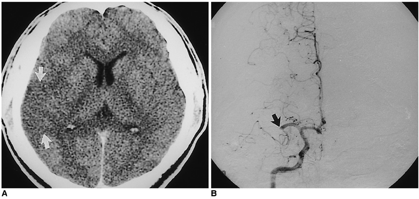
Fig. 2 A 32-year-old man who reported the onset of hemiplegia three hours prior to examination (case 2). A. Contrast-enhanced CT scan shows subtle low density in the right temporal lobe and insular cortex(arrows), suggesting a hyper-acute infarct in the territory of the right middle cerebral artery. B. Frontal view of right internal carotid angiogram shows complete occlusion of the right middle cerebral artery at M1 portion(arrow).

Fig. 3 A 34-year-old woman with progressive dementia (case 8). Axial T1-weighed MR image shows diffuse cerebral atrophy with prominent sulci and dilatation of the lateral ventricles.
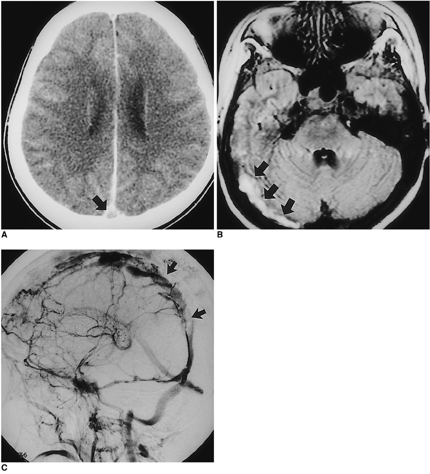
Fig. 4 A 25-year-old woman who had suffered right hemiplegia for 2 days (case 6). A. Enhanced CT scan shows a filling defect in the superior sagittal sinus (empty delta sign, arrow). B. FLAIR axial T2-weighted MR image shows abnormally high signal intensity in the right transverse sinus, suggesting thrombosis (arrows). C. Oblique view of right internal carotid angiogram during the venous phase shows multiple filling defects in the superior sagittal sinus (arrows).
Reference
-
1. Harris EN, Gharavi AE, Hughes GRV. Antiphospholipid antibodies. Clin Rheum Dis. 1985. 11:591–609.2. Harris EN, Baguley E, Asherson RA, et al. Clinical and serological features of the "antiphospholipid syndrome". Br J Rheumatol. 1987. 26:S. 19.3. Hughson MD, Mccarty GA, Brumback RA. Spectrum of vascular pathology affecting patients with the antiphospholipid syndrome. Hum Pathol. 1995. 26:716–724.4. Asherson RA, Khamashta MA, Ordi-Ros J, et al. The "primary" antiphospholiupid syndrome: major clinical and serological features. Medicine. 1989. 68:366–374.5. Gharavi AE, Wilson WA. The syndrome of thrombosis, thrombocytopenia, and recurrent spontaneous abortions associated with antiphospholipid antibodies: Hughes syndrome. Lupus. 1996. 5:343–344.6. Provenzale JM, Heinz ER, Ortel TL, et al. Antiphospholipid antibodies in patients without SLE: neuroradiologic findings. Radiology. 1994. 192:531–537.7. Provenzale JM, Barboriak DP, Allen NB, Ortel TL. Patients with antiphospholipd antibodies: CT and MR findings of the brain. AJR. 1996. 167:1573–1578.8. Toubi E, Khamashta MA, Panarra A, Hugham GRV. Association of antiphospholipid antibodies with central nervous system disease in SLE. Am J Med. 1995. 99:397–401.9. Brey RL, Gharavi AE, Lockshin MD. Neurologic complications of antiphospholipid antibodies. Rheumatol Clin North Am. 1993. 4:833–850.10. Oeffinger KC, Roaten SP. Antihpospholipid syndrome. J Fam Prac. 1994. 38:611–619.11. Petri M. Pathogenesis and treatment of the antiphospholipid antibody syndrome. Med Clin North Am. 1997. 81:151–177.12. Aron AL, Gharavi AE, Shoenfeld Y. Mechanisms of action of antiphospholipid antibodies in the antiphospholipid antibodies in the antiphospholipd syndrome. Int Arch Allergy Immunol. 1995. 106:8–12.13. Lechner K, Pabinger-Fasching I. Lupus anticoagulants and thrombosis: a study of 25 cases and review of the literature. Haemostasis. 1985. 15:254–262.14. Levine SR, Brey RL. Neurological aspects of antihpospholipid antibody syndrome. Lupus. 1996. 5:347–353.15. Westerman EM, Miles JM. Neuropathologic findings in multi-infarct dementia associated with anticardiolipin antibody. Arthritis Rheum. 1992. 35:1038–1041.16. Coull BM, Bourdette DN, Goodnight SH, Briley DP, Hart R. Multiple cerebral infarctions and dementia as associated with anticardiolipin antibodies. Stroke. 1987. 18:1107–1112.17. Westerman EM, Miles KM, Backonja M, Sundstrom WR. Neuropahtologic findings in multi-infarct dementia associated with anticardiolipin antibody. Arthritis Rheum. 1992. 9:1038–1041.18. Levin SR, Kieran S, Puzio K, et al. Cerebral venous thrombosis with lupus anticoagulants: report of two cases. Stroke. 1987. 18:801–804.19. Carhuapoma JR, Mitsias P, Levine SR. Cerebral venous thrombosis and antiphospholipid antibodies. Stroke. 1997. 28:2363–2369.
- Full Text Links
-
- Actions
-
Cited
- CITED
-
- Close
- Share
-
- Similar articles
-
- A patient with chorea associated with hyperthyroidism and primary antiphospholipid antibody syndrome
- A Case of Primary Adrenal Insufficiency Presenting as the Initial Clinical Manifestation of Primary Antiphospholipid Antibody Syndrome
- A Case of Refractory Headache with Antiphospholipid Antibody Syndrome Improved by High-Intensity Warfarin Medication
- Flank ulcer in a patient with primary antiphospholipid syndrome
- Antiphospholipid Antibody Syndrome
