Pure Epidural Cavernous Hemangioma of the Cervical Spine that Presented with an Acute Sensory Deficit Caused by Hemorrhage
- Affiliations
-
- 1Departmet of Diagnostic Radiology, Wooridul Spine Hospital, Seoul, Korea. jbj135@hanafos.com
- 2Pochon CHA University, Bundang CHA Hospital, Sungnam-si, Korea.
- 3Yongdong Severance Hospital, Yonsei University College of Medicine, Seoul, Korea.
- 4Departmet of Neurosurgery, Wooridul Spine Hospital, Seoul, Korea.
- 5Departmet of Pathology, Wooridul Spine Hospital, Seoul, Korea.
- 6T and C Pathology Hospital, Seoul, Korea.
- KMID: 1777180
- DOI: http://doi.org/10.3349/ymj.2006.47.6.877
Abstract
- Pure epidural cavernous hemangioma of the spine without vertebral involvement is rare. Due to the slow growth of this lesion, the most common symptoms are chronic pain, myelopathy, and radiculopathy. In our case, the patient complained of an acute onset sensory deficit of the C4 dermatome. An MRI revealed an epidural mass with an acute hematoma. Here, we report a case of a pure epidural cavernous hemangioma that presented with acute neurologic symptoms caused by intralesional hemorrhage and an acute epidural hematoma, which were demonstrated on the patient's MRI.
MeSH Terms
-
Tomography, X-Ray Computed
Middle Aged
Male
Hyperesthesia/*diagnosis/etiology
Humans
Hematoma, Epidural, Spinal/complications/*diagnosis/radiography
Hemangioma, Cavernous, Central Nervous System/complications/*diagnosis/radiography
Epidural Space/radiography
Epidural Neoplasms/complications/*diagnosis/radiography
Cervical Vertebrae
Figure
-
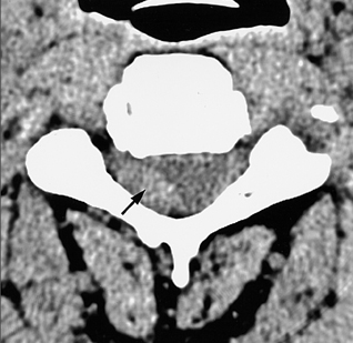
Fig. 1 Axial CT image shows slightly hyperdense, oval-shaped mass (arrow) anterior to the right lamina.
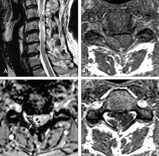
Fig. 2 (A) Sagittal T2-weighted image shows a posterior epidural mass with peripheral high signal intensity (white arrowheads) and a central low signal area (black arrow). The mass is well-demarcated from the spinal cord by a dark signal line representing the dura. The triangular shaped epidural lesion above and below the main lesion shows high signal intensity (white arrow). This area was later confirmed as a hyperacute hematoma in the epidural space. (B) This lesion was isodense with spinal cord on axial T1-weighted image. (C) Axial gradient-echo (fast field gradient echo, TR/TE/FA, 536/23/25 degree) image shows very dark central signal area and a peripheral dark signal rim similar to the area in the peripheral portion of the mass (white arrow). Initially, this area was thought to be a hemosiderin rim; however, pathological findings showed it be an acute hematoma. This dark signal characteristic of the gradient-echo image may be caused by deoxy-hemoglobin in the hematoma. The remaining mass shows high signal intensity (black arrow). Outside of the main lesion is a bright signal epidural lesion (black arrowheads) that was confirmed to be a hyperacute hematoma in the epidural space. (D) After Gadolinium-DTPA contrast enhancement, the mass shows peripheral heterogeneous enhancement (white arrow). Surrounding epidural hyperacute hematoma shows strong contrast enhancement (white arrowheads).
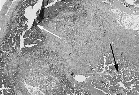
Fig. 3 Photomicrograph shows a cavernous hemanigoma composed of thin-walled vascular channels in collagenous tissue. There was a large central hematoma (black arrow) and peripheral hematoma (white arrow) (H&E stain, ×40). There was no hemosiderin deposition in the mass.
Cited by 2 articles
-
Spinal Cord Tumors of the Thoracolumbar Junction Requiring Surgery: A Retrospective Review of Clinical Features and Surgical Outcome
Dong Ah Shin, Sang Hyun Kim, Keung Nyun Kim, Hyun Cheol Shin, Do Heum Yoon
Yonsei Med J. 2007;48(6):988-993. doi: 10.3349/ymj.2007.48.6.988.Spinal Epidural Arteriovenous Hemangioma Mimicking Lumbar Disc Herniation
Kyung Hyun Kim, Sang Woo Song, Soo Eon Lee, Sang Hyung Lee
J Korean Neurosurg Soc. 2012;52(4):407-409. doi: 10.3340/jkns.2012.52.4.407.
Reference
-
1. Graziani N, Bouillot P, Figarella-Branger D, Dufour H, Peragut JC, Grisoli F. Cavernous angiomas and arteriovenous malformations of the spinal epidural space: report of 11 cases. Neurosurgery. 1994. 35:856–864.2. Tekkok IH, Akpinar G, Gungen Y. Extradural lumbosacral cavernous hemangioma. Eur Spine J. 2004. 13:469–473.3. Rovira A, Rovira A, Capellades J, Zauner M, Bella R, Rovira M. Lumbar extradural hemangioms: reports of three cases. AJNR Am J Neuroradiol. 1999. 20:27–31.4. Goyal A, Singh AK, Gupta V, Tatke M. Spinal epidural cavernous hemangioma: a case report and review of literature. Spinal Cord. 2002. 40:200–202.5. Talacchi A, Spinnato S, Alessandrini F, Iuzzolino P, Bricolo A. Radiologic and surgical aspects of pure spinal epidural cavernous angiomas. Report on 5 cases and review of the literature. Surg Neurol. 1999. 52:198–203.6. Shin JH, Lee HK, Rhim SC, Park SH, Choi CG, Suh DC. Spinal epidural cavernous hemangioma: MR fingings. J Comput Assist Tomogr. 2001. 25:257–261.7. Gupta S, Kumar S, Banerji D, Pandey R, Gujral R. Magnetic resonance imaging features of an epidural spinal haemangioma. Australas Radiol. 1996. 40:342–344.8. Carlier R, Engerand S, Lamer S, Vallee C, Bussel B, Polivka M. Foraminal epidural extra osseous cavernous hemangioma of the cervical spine: a case report. Spine. 2000. 25:629–631.9. Zevgaridis D, Buttner A, Weis S, Hamburger C, Reulen HJ. Spinal epidural cavernous hemangiomas. Report of three cases and review of the literature. J Neurosurg. 1998. 88:903–908.10. Groen RJ, Ponssen H. The spontaneous spinal epidural hematoma: A study of the etiology. J Neurol Sci. 1990. 98:121–138.11. Kubo Y, Nishiura I, Koyama T. Repeated transient paraparesis due to solitary spinal epidural arteriovenous malformation- a case report. No Shinkei Geka. 1984. 12:857–862.12. Graziani N, Bouillot P, Figarella-Branger D, Dufour H, Peragut JC, Grisoli F. Cavernous angiomas and arteriovenous malformations of the spinal epidural space: report of 11 cases. Neurosurgery. 1994. 35:856–863.13. Miyagi Y, Miyazono M, Kamikaseda K. Spinal epidural vascular malformation presenting in association with a spontaneously resolved acute epidural hematoma. Case report. J Neurosurg. 1998. 88:909–911.14. Hentschel SJ, Woolfenden AR, Fairholm DJ. Resolution of spontaneous spinal epidural hematoma without surgery: report of two cases. Spine. 2001. 26:E525–E527.
- Full Text Links
-
- Actions
-
Cited
- CITED
-
- Close
- Share
-
- Similar articles
-
- Two Cases of Epidural Cavernous Hemangioma in the Thoraic Spine
- Pure Spinal Epidural Cavernous Hemangioma with Intralesional Hemorrhage: A Rare Cause of Thoracic Myelopathy
- Pure Epidural Cavernous Hemangioma in Thoracic Region: A Case Report
- Spinal Epidural Cavernous Hemangioma Simulating a Disc Protrusion: A Case Report
- Pure Thoracic Spinal Epidural Cavernous Hemangioma with Spinal Cord Compression: A Case Report
