Changes of Functional MRI Findings in a Patient Whose Pathological Gambling Improved with Fluvoxamine
- Affiliations
-
- 1Department of Psychiatry, Chonbuk National University Medical School, Jeonju, Korea.
- 2Department of Diagnostic Radiology, Mirae Hospital, Gimje, Korea.
- 3Department of Diagnostic Radiology, Shinsegae Hospital, Gimje, Korea.
- 4Department of Diagnostic Radiology, Chonbuk National University Medical School, Jeonju, Korea.
- 5Department of Psychiatry, Sungkyunkwan University School of Medicine, Kangbuk Samsung Hospital, Seoul, Korea.
- 6Department of Psychiatry, College of Medicine, Catholic University of Korea, Bucheon, Korea.
- 7Department of Psychiatry, University of Ulsan College of Medicine, Ulsan University Hospital, Ulsan, Korea. peaceinu@yahoo.co.kr
- KMID: 1758575
- DOI: http://doi.org/10.3349/ymj.2009.50.3.441
Abstract
- Legalized gambling is a growing industry, and is probably a factor in the presently increasing prevalence of pathological gambling. We present a case of a 36-year-old pathological gambler who was treated with fluvoxamine, a selective serotonin reuptake inhibitor, and who was assessed by functional MRI before and after drug administration. During activation periods, the pathological gambler was shown cards as stimuli, and fMRI results in several brain regions showed differential effects before and after medication and a maintenance period. This case demonstrates that the treatment response to fluvoxamine in a pathological gambler was observed not only by subjective self-report, but also by objective fMRI results. Therefore, fMRI may be a useful tool in the diagnosis and prediction of treatment response in patients afflicted with pathological gambling.
Keyword
MeSH Terms
Figure
-
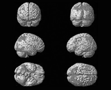
Fig. 1 fMRI before fluvoxamine administration. This figure shows the difference between the activation and resting conditions. The brain is generally activated, especially in both parietal regions and the occipital, and frontal regions.
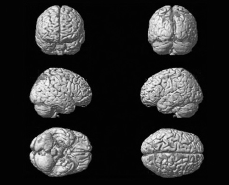
Fig. 2 fMRI one week after fluvoxamine administration. The figure shows the difference between the activation and resting conditions. There is no activated region in the occipital and temporal areas. There were activated regions in both frontal (right: Brodmann's areas 6, 5, and 4; left: Brodmann's areas 6, 4) and both parietal (right: Brodmann's area 7; left: Brodmann's areas 7, 19) lobes.

Fig. 3 fMRI six weeks after fluvoxamine administration. The figure shows the difference between the activation and resting conditions. There were activated regions in the left frontal (Brodmann's area 6), right parietal (Brodmann's areas 7, 40), and left parietal (Brodmann's areas 7, 19, and 40) lobes.
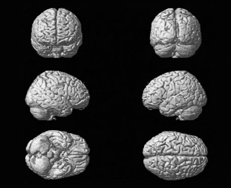
Fig. 4 fMRI six months after fluvoxamine administration. The figure shows the difference between the activation and resting conditions. The previously activated regions were generally decreased. There were small activated regions in the right frontal lobe (Brodmann's area 6), left occipital lobe (Brodmann's areas 18, 19), right limbic area (Brodmann's area 24), and left limbic area (Brodmann's area 31).
Reference
-
1. Potenza MN, Kosten TR, Rounsaville BJ. Pathological gambling. JAMA. 2001. 286:141–144.
Article2. Blanco C, Moreyra P, Nunes EV, Sáiz-Ruiz J, Ibánez Ä. Pathological gambling: addiction or compulsion? Semin Clin Neuropsychiatry. 2001. 6:167–176.
Article3. Potenza MN. The neurobiology of pathological gambling. Semin Clin Neuropsychiatry. 2001. 6:217–226.
Article4. Petry NM, Armentano C. Prevalence, assessment, and treatment of pathological gambling: a review. Psychiatr Serv. 1999. 50:1021–1027.
Article5. Hollander E, DeCaria CM, Finkell JN, Begaz T, Wong CM, Cartwright C. A randomized double-blind fluvoxamine/placebo crossover trial in pathologic gambling. Biol Psychiatry. 2000. 47:813–817.
Article6. Potenza MN, Leung HC, Blumberg HP, Peterson BS, Fulbright RK, Lacadie CM, et al. An FMRI Stroop task study of ventromedial prefrontal cortical function in pathological gamblers. Am J Psychiatry. 2003. 160:1990–1994.7. Choi WC, Kim KB, Oh DY, Lee TK. A preliminary Study on standardization of Korean form of South Oak Gambling Screening. J Korean Acad Addict Psychiatry. 2001. 5:46–52.8. Pallanti S, Bernardi S, Quercioli L, DeCaria C, Hollander E. Serotonin dysfunction in pathological gamblers: increased prolactin response to oral m-CPP versus placebo. CNS Spectr. 2006. 11:956–964.9. Hollander E, Buchalter AJ, DeCaria CM. Pathological gambling. Psychiatr Clin North Am. 2000. 23:629–642.
Article10. Blanco C, Orensanz-Muñoz L, Blanco-Jerez C, Saiz-Ruiz J. Pathological gambling and platelet MAO activity: a psychobiological study. Am J Psychiatry. 1996. 153:119–121.11. Bechara A, Damasio AR, Damasio H, Anderson SW. Insensitivity to future consequences following damage to human prefrontal cortex. Cognition. 1994. 50:7–15.
Article12. Stein EA, Pankiewicz J, Harsch HH, Cho JK, Fuller SA, Hoffmann RG, et al. Nicotine-induced limbic cortical activation in the human brain: a functional MRI study. Am J Psychiatry. 1998. 155:1009–1015.
Article13. Breiter HC, Gollub RL, Weisskoff RM, Kennedy DN, Makris N, Berke JD, et al. Acute effects of cocaine on human brain activity and emotion. Neuron. 1997. 19:591–611.
Article14. Potenza MN, Fiellin DA, Heninger GR, Rounsaville BJ, Mazure CM. Gambling: an addictive behavior with health and primary care implications. J Gen Intern Med. 2002. 17:721–732.15. Saxena S, Rauch SL. Functional neuroimaging and the neuroanatomy of obsessive compulsive disorder. Psychiatr Clin North Am. 2000. 23:563–586.
Article
- Full Text Links
-
- Actions
-
Cited
- CITED
-
- Close
- Share
-
- Similar articles
-
- Association Between Pathological Gambling and Depression in Korean Adults
- Stress, Coping Style and Depression in Pathological Gamblers
- Factors Influencing Recognition of Motivation for Change in Pathological Gamblers
- Factors Influencing Problem and Pathological Gambling in Participants of Horse Race Gambling
- A Case of Frontotemporal Dementia with Amyotrophic Lateral Sclerosis Presenting with Pathological Gambling
