Nodule Detection in a Lung Region that's Segmented with Using Genetic Cellular Neural Networks and 3D Template Matching with Fuzzy Rule Based Thresholding
- Affiliations
-
- 1Istanbul Commerce University, Ragip Gumuspala Cad. No: 84 34378 Eminonu, Istanbul, Turkey. serhat@iticu.edu.tr
- 2Istanbul University, Engineering Faculty, Electrical and Electronics Eng. Dept., 34850, Avcilar, Istanbul, Turkey.
- KMID: 1734269
- DOI: http://doi.org/10.3348/kjr.2008.9.1.1
Abstract
OBJECTIVE
The purpose of this study was to develop a new method for automated lung nodule detection in serial section CT images with using the characteristics of the 3D appearance of the nodules that distinguish themselves from the vessels. MATERIALS AND METHODS: Lung nodules were detected in four steps. First, to reduce the number of region of interests (ROIs) and the computation time, the lung regions of the CTs were segmented using Genetic Cellular Neural Networks (G-CNN). Then, for each lung region, ROIs were specified with using the 8 directional search; +1 or -1 values were assigned to each voxel. The 3D ROI image was obtained by combining all the 2-Dimensional (2D) ROI images. A 3D template was created to find the nodule-like structures on the 3D ROI image. Convolution of the 3D ROI image with the proposed template strengthens the shapes that are similar to those of the template and it weakens the other ones. Finally, fuzzy rule based thresholding was applied and the ROI's were found. To test the system's efficiency, we used 16 cases with a total of 425 slices, which were taken from the Lung Image Database Consortium (LIDC) dataset. RESULTS: The computer aided diagnosis (CAD) system achieved 100% sensitivity with 13.375 FPs per case when the nodule thickness was greater than or equal to 5.625 mm. CONCLUSION: Our results indicate that the detection performance of our algorithm is satisfactory, and this may well improve the performance of computer-aided detection of lung nodules.
Keyword
MeSH Terms
Figure
-

Fig. 1 Procedural flowchart for detecting lung nodules.

Fig. 2 A. First CT image, B. Segmented lung region using cellular neural network, C. CT image in the lung region, D. Voxels having suitable density values, E. ROIs in the lung region, F. Detected nodule region.
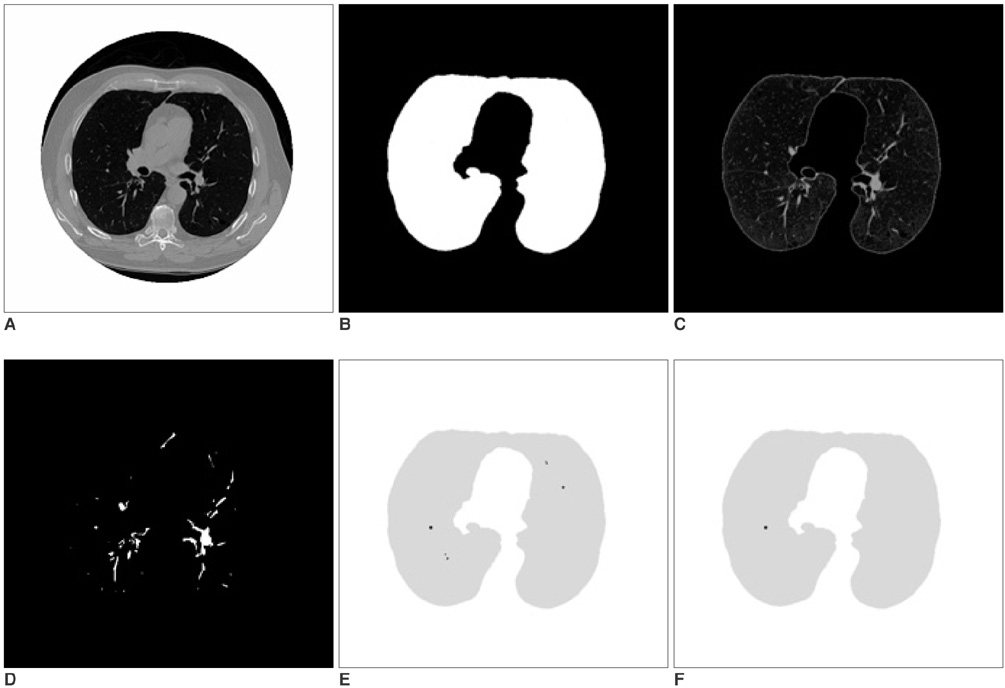
Fig. 3 A. Second CT image, B. Segmented lung region using cellular neural network, C. CT image in the lung region, D. Voxels having suitable density values, E. ROIs in the lung region, F. Detected nodule region.
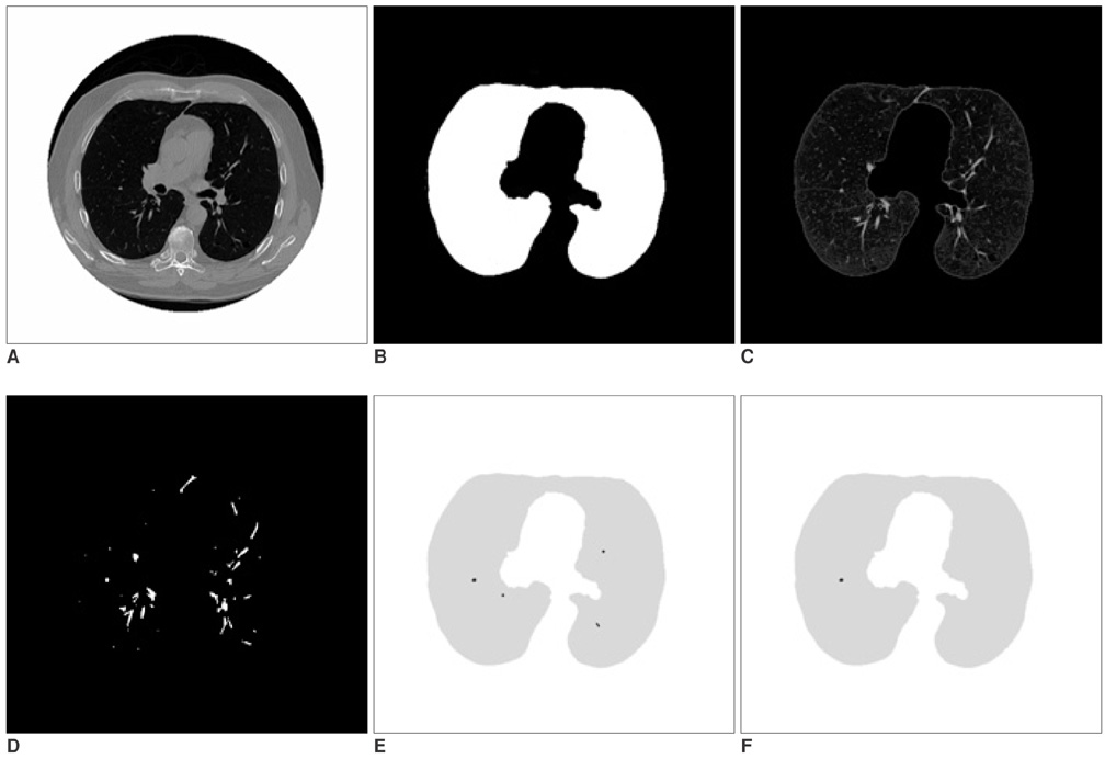
Fig. 4 A. Third CT image, B. Segmented lung region using cellular neural network, C. CT image in the lung region, D. Voxels having suitable density values, E. ROIs in the lung region, F. Detected nodule region.
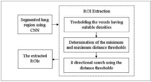
Fig. 5 Procedural flowchart of ROI specification.
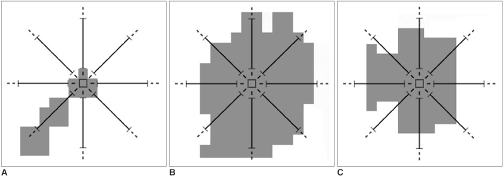
Fig. 6 A. A voxel which doesn't have a number of adjacent neighbor voxels greater than or equal to the value of "minimum distance threshold", so it is not a part of the ROI. B. A voxel which doesn't have a number of adjacent neighbor voxels less than or equal to the value of "maximum distance threshold", so it is not a part of the ROI. C. A voxel which has a number of adjacent neighbor voxels greater than or equal to the value of "minimum distance threshold", and less than or equal to the value of "maximum distance threshold", so it is a part of the ROI.
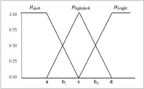
Fig. 7 Threshold membership functions
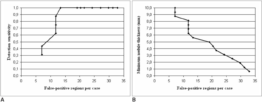
Fig. 8 A. Free-response receiver operating characteristic curve showing the performance of the nodule detection task according to the numbers of cases. B. Minimum nodule thickness versus false-positive regions per case.
Cited by 1 articles
-
An Engineering View on Megatrends in Radiology: Digitization to Quantitative Tools of Medicine
Namkug Kim, Jaesoon Choi, Jaeyoun Yi, Seungwook Choi, Seyoun Park, Yongjun Chang, Joon Beom Seo
Korean J Radiol. 2013;14(2):139-153. doi: 10.3348/kjr.2013.14.2.139.
Reference
-
1. Greenlee RT, Murray T, Bolden S, Wingo PA. Cancer statistics, 2000. CA Cancer J Clin. 2000. 50:7–33.2. Giger ML, Bae KT, MacMahon H. Computerized detection of pulmonary nodules in computed tomography images. Invest Radiol. 1994. 29:459–465.3. Armato SG 3rd, Giger ML, Moran CJ, Blackburn JT, Doi K, MacMahon H. Computerized detection of pulmonary nodules on CT scans. Radiographics. 1999. 1303–1311.4. Kanazawa K, Kawata Y, Niki N, Satoh H, Ohmatsu H, Kakinuma R. Doi K, MacMahon H, Giger ML, Hoffmann K, editors. Computer-aided diagnostic system for pulmonary nodules based on helical CT images. Computer-aided diagnosis in medical imaging. 1998. . Amsterdam, the Netherlands: Elsevier Science;131–136.5. Penedo MG, Carreira MJ, Mosquera A, Cabello D. Computer-aided diagnosis: a neural-network-based approach to lung nodule detection. IEEE Trans Med Imaging. 1998. 17:872–880.6. Xu XW, Doi K, Kobayashi T, MacMahon H, Giger ML. Development of an improved CAD scheme for automated detection of lung nodules in digital chest images. Med Phys. 1997. 24:1395–1403.7. Lo SCB, Lou SLA, Lin JS, Freedman M, Chien MV, Mun SK. Artificial convolution neural network techniques and applications for lung nodule detection. IEEE Trans Med Imaging. 1995. 14:711–718.8. Brown MS, McNitt-Gray MF, Goldin JG, Suh RD, Sayre JW, Aberle DR. Patient-specific models for lung nodule detection and surveillance in CT images. IEEE Trans Med Imaging. 2001. 20:1242–1250.9. Betke M, Hong H, Thomas D, Prince C, Ko JP. Landmark detection in the chest and registration of lung surfaces with an application to nodule registration. Med Image Anal. 2003. 7:265–281.10. Lee Y, Hara T, Fujita H, Itoh S, Ishigaki T. Automated detection of pulmonary nodules in helical CT images based on an improved template-matching technique. IEEE Trans Med Imaging. 2001. 20:595–604.11. Farag AA, El-Baz A, Gimel'farb G, Falk R. Detection and recognition of lung abnormalities using deformable templates. Proceedings of the 17th International Conference on Pattern Recognition. 2004. 3:738–741.12. Zadeh LA. Fuzzy Sets. Information and Control. 1965. 8:338–353.13. Armato SG 3rd, McLennan G, McNitt-Gray MF, Meyer CR, Yankelevitz D, Aberle DR, et al. Lung image database consortium: developing a resource for the medical imaging research community. Radiology. 2004. 232:739–748.14. Hounsfield GN. Novel award address. Computed medical imaging. Med Phys. 1980. 7:283–290.15. Chua LO, Yang L. Cellular neural networks: theory. IEEE Trans Circuits and Syst. 1988. 35:1257–1272.16. Holland JH. Adaptation in neural and artificial systems. 1975. Ann Arbor, MI: University of the Michigan Press.17. Kozek T, Roska T, Chua LO. Genetic algorithms for CNN template learning. IEEE Trans Circuit and Syst. 1988. 40:392–402.18. Davis L. Handbook of genetic algorithms. 1991. New York: Van Nostrand Reinhold.19. Ozekes S, Osman O, Camurcu AY. Mammographic mass detection using a mass template. Korean J Radiol. 2005. 6:221–228.20. Ozekes S, Camurcu AY. Automatic lung nodule detection using template matching. Lecture Notes in Computer Science. 2006. 4243:247–253.21. Cheng HD, Chen JR, Li J. Threshold selection based on fuzzy cpartition entropy approach. Pattern Recognition. 1998. 31:857–870.22. Armato SG 3rd, Sensakovic WF. Automated lung segmentation for thoracic CT impact on computer-aided diagnosis. Acad Radiol. 2004. 11:1011–1021.23. Brown MS, McNitt-Gray MF, Mankovich NJ, Goldin JG, Hiller J, Wilson LS, et al. Method for segmenting chest CT image data using an anatomical model: preliminary results. IEEE Trans Med Imaging. 1997. 16:828–839.24. Hu S, Hoffman EA, Reinhardt JM. Automatic lung segmentation for accurate quantitation of volumetric X-ray CT images. IEEE Trans Med Imaging. 2001. 20:490–498.25. Silva A, Silva JS, Santos BS, Ferreira C. Fast pulmonary contour extraction in X-ray CT images: a methodology and quality assessment. Proc SPIE. 2001. 4321:216–224.26. Zheng B, Leader JK, Maitz GS, Chapman BE, Fuhrman CR, Rogers RM, et al. A simple method for automated lung segmentation in X-ray CT images. Proc SPIE (Medical Imaging). 2003. 5032:1455–1146.27. Leader JK, Zheng B, Rogers RM, Sciurba FC, Perez A, Chapman BE, et al. Automated lung segmentation in X-ray computed tomography: development and evaluation of heuristic threshold-based scheme. Acad Radiol. 2003. 10:1224–1236.28. Kakeda S, Moriya J, Sato H, Aoki T, Watanabe H, Nakata H, et al. Improved detection of lung nodules on chest radiographs using a commercial computer-aided diagnosis system. AJR Am J Roentgenol. 2004. 182:505–510.29. Wood SA, Stapleton SJ, Schneider AC, Carano S, Herold CJ, Castellino RA. Computer-aided detection (CAD) of actionable lung nodules on multi-slice CT (MSCT) scans of the lung: sensitivity and false positive marker rates. Radiology. 2002. 225:477.
- Full Text Links
-
- Actions
-
Cited
- CITED
-
- Close
- Share
-
- Similar articles
-
- A study on Recognition of Bronchogenic Cancer Cell using Fuzzy Neural Networks
- 3D Visualization of 2D CT Bone Image Using FSTB Algorithm
- Evaluation of Semi-automatic Segmentation Methods for Persistent Ground Glass Nodules on Thin-Section CT Scans
- Manufacture of the Serially Sectioned Images of the Whole Body (Fifth Report: Methods for Manufacture of the Three Dimensional Images and Virtual Dissection Software)
- Adaptive Thresholding for Pap-Smear
