An outbreak of fatal hemorrhagic pneumonia caused by Streptococcus equi subsp. zooepidemicus in shelter dogs
- Affiliations
-
- 1Animal Disease Diagnostic Center, National Veterinary Research and Quarantine Service, Anyang 430-824, Korea. jwbyun@nvrqs.go.kr
- KMID: 1726929
- DOI: http://doi.org/10.4142/jvs.2009.10.3.269
Abstract
- An outbreak of fatal hemorrhagic pneumonia with 70~90% morbidity and 50% mortality occurred in an animal shelter in Yangju, Gyeonggi Province, Korea. Clinically, the affected dogs showed severe respiratory distress within 48 h after arriving in the shelter. The dead were found mainly with nasal bleeding and hematemesis. At necropsy, hemothorax and hemorrhagic pneumonia along with severe pulmonary consolidation was observed, though histopathological analysis showed mainly hemorrhagic bronchopneumonia. Lymphoid depletion was inconsistently seen in the spleen, tonsil and bronchial lymph node. Gram-positive colonies were shown in blood vessels or parenchyma of cerebrum, lung, liver, spleen, and kidney. Also, Streptococcus (S.) equi subsp. zooepidemicus was isolated from the various organs in which the bacterium was microscopically and histologically detected. In addition, approximately 0.9 Kb specific amplicon, antiphagocytic factor H binding protein, was amplified in the bacterial isolates. In this study, we reported an outbreak of canine hemorrhagic bronchopneumonia caused by S. equi subsp. zooepidemicus in an animal shelter in Yangju, Korea.
MeSH Terms
Figure
-

Fig. 1 (A) Lung. There is severe alveolar congestion, with a mixture of edema fluids, inflammatory cells and erythrocytes infiltrating the alveolar cavity and bronchiole. H&E stain. Scale bar = 200 µm. (B) Lung. Gram-positive cocci are scattered in alveolar lumen and also engulfed by alveolar macrophages (arrows). Gram stain. Scale bar = 20 µm. (C) Liver. Bacterial clumps are infiltrated in the sinusoid. Gram stain. Scale bar = 50 µm. (D) Cerebrum. Lots of cocci are mixed with red blood cells and monocytes in a meningeal blood vessel. Gram stain. Scale bars = 50 µm.
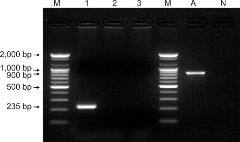
Fig. 2 Amplified products of Streptococcus zooepidemicus isolated from the lungs of dogs with hemorrhagic pneumonia. Lane M: DNA size marker (100 bp ladder), Lane 1-3: Amplified products for sodA, seeH and seeI, respectively. Lane A: DNA fragment using primer for FUS and FDS located upstream and downstream of se18.9 from the isolated bacteria. Lane N: Negative control.
Reference
-
1. Alber J, El-Sayed A, Estoepangestie S, Lämmler C, Zschöck M. Dissemination of the superantigen encoding genes seeL, seeM, szeL and szeM in Streptococcus equi subsp. equi and Streptococcus equi subsp. zooepidemicus. Vet Microbiol. 2005. 109:135–141.2. Alber J, El-Sayed A, Lämmler C, Hassan AA, Weiss R, Zschöck M. Multiplex polymerase chain reaction for identification and differentiation of Streptococcus equi subsp. zooepidemicus and Streptococcus equi subsp. equi. J Vet Med B Infect Dis Vet Public Health. 2004. 51:455–458.
Article3. Båverud V, Johansson SK, Aspan A. Real-time PCR for detection and differentiation of Streptococcus equi subsp. Equi and Streptococcus equi subsp. zooepidemicus. Vet Microbiol. 2007. 124:219–229.
Article4. Chalker VJ, Brooks HW, Brownlie J. The association of Streptococcus equi subsp. zooepidemicus with canine infectious respiratory disease. Vet Microbiol. 2003. 95:149–156.
Article5. Elia G, Decaro N, Martella V, Cirone F, Lucente MS, Lorusso E, Di Trani L, Buonavoglia C. Detection of canine distemper virus in dogs by real-time RT-PCR. J Virol Methods. 2006. 136:171–176.
Article6. Hu RL, Huang G, Qiu W, Zhong ZH, Xia XZ, Yin Z. Detection and differentiation of CAV-1 and CAV-2 by polymerase chain reaction. Vet Res Commun. 2001. 25:77–84.7. Garnett NL, Eydelloth RS, Swindle MM, Vonderfecht SL, Strandberg JD, Luzarraga MB. Hemorrhagic streptococcal pneumonia in newly procured research dogs. J Am Vet Med Assoc. 1982. 181:1371–1374.8. Greene CE. Infectious Diseases of the Dog and Cat. 2006. 3rd ed. Philadelphia: Saunders;302–309.9. Kim MK, Jee H, Shin SW, Lee BC, Pakhrin B, Yoo HS, Yoon JH, Kim DY. Outbreak and control of haemorrhagic pneumonia due to Streptococcus equi subspecies zooepidemicus in dogs. Vet Rec. 2007. 161:528–530.10. Ladlow J, Scase T, Waller A. Canine strangles case reveals a new host susceptible to infection with Streptococcus equi. J Clin Microbiol. 2006. 44:2664–2665.
Article11. Pesavento PA, Hurley KF, Bannasch MJ, Artiushin S, Timoney JFA. A clonal outbreak of acute fatal hemorrhagic pneumonia in intensively housed (shelter) dogs caused by Streptococcus equi subsp. zooepidemicus. Vet Pathol. 2008. 45:51–53.
Article12. Tiwari R, Qin A, Artiushin S, Timoney JF. Se18.9, an anti-phagocytic factor H binding protein of Streptococcus equi. Vet Microbiol. 2007. 121:105–115.13. Walker JA, Timoney JF. Molecular basis of variation in protective SzP proteins of Streptococcus zooepidemicus. Am J Vet Res. 1998. 59:1129–1133.
- Full Text Links
-
- Actions
-
Cited
- CITED
-
- Close
- Share
-
- Similar articles
-
- Persistent occurrence of a single Streptococcus equi subsp. zooepidemicus clone in the pig and monkey population in Indonesia
- Etiologic and epidemiologic analysis of bacterial infectious upper respiratory disease in Thoroughbred horses at the Seoul Race Park
- Fatal pneumonia caused by extraintestinal pathogenic Escherichia coli in a young dog
- Group B Streptococcal Toxic Shock-like Syndrome: A Case Report and Review of the Literature
- A Case of Streptococcal Toxic Shock Syndrome Caused by Group A Streptococcus Pneumonia
