Utility of Optical Coherence Tomography to Assess a Hazy Intracoronary Image after Percutaneous Coronary Intervention
- Affiliations
-
- 1University Hospital La Paz, Interventional Cardiology Department, Madrid, Spain. sebastian_carrizo@live.com
- KMID: 1723000
- DOI: http://doi.org/10.4070/kcj.2013.43.1.44
Abstract
- Although its use in daily practice is not common, optical coherence tomography (OCT) is a powerful research tool in invasive cardiology. This report describes a hazy angiography image after percutaneous coronary intervention that has been assessed using OCT. Based on the results of the OCT, the patient underwent an elective coronary angioplasty with standard anticoagulation. After implantation of the stent, an intracoronary hazy image was seen on angiography. The use of OCT permitted a correct diagnosis and a successful treatment. This paper provides a discussion of the advantages and disadvantages of OCT, and a comparison with intravascular ultrasound.
Keyword
MeSH Terms
Figure
-
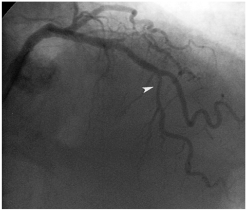
Fig. 1 A severe lesion in the middle portion of the left anterior descendent coronary artery (arrow head).
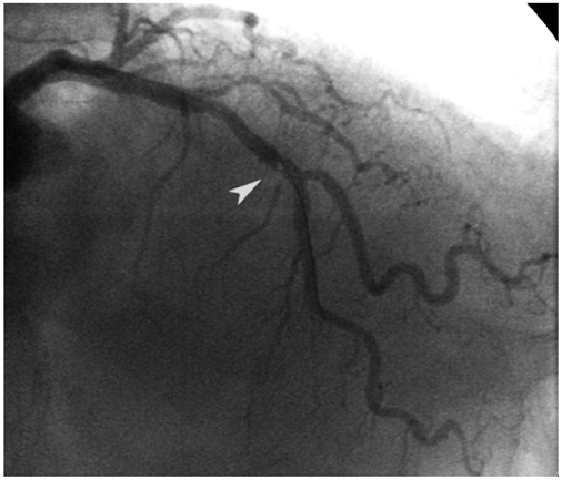
Fig. 2 Intrastent luminal hazy image on the middle portion of the left anterior descendent coronary artery (arrow head).
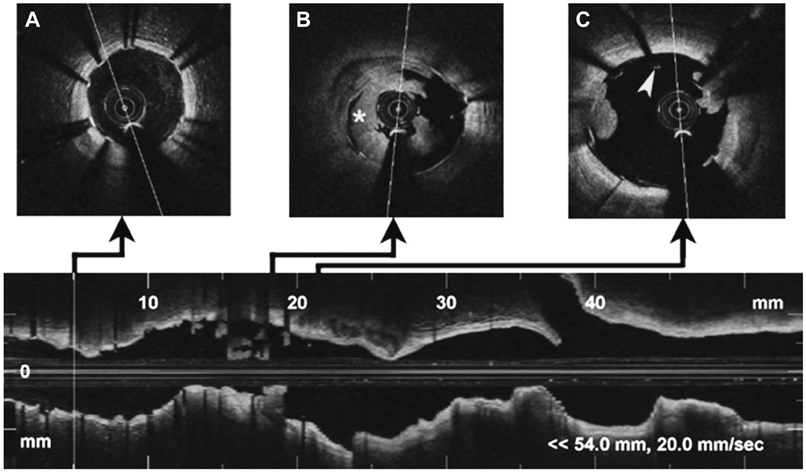
Fig. 3 Optical coherence tomography imaging of left anterior descending artery. Distal (A): correct apposition of the stent without intraluminal content. Middle (B): a signal rich, low-backscattering protrusion image compatible with white thrombus (mark), which occupies the greater part of the vessel lumen. Proximal (C): stent malapposition (arrow head) in the proximal border, with small images of thrombus.
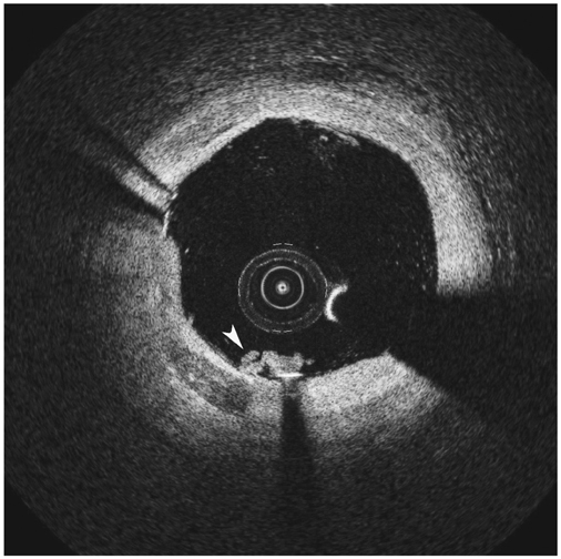
Fig. 4 Final optical coherence tomography imaging of the left anterior descending artery. There is almost complete reduction of the thrombus (arrow head) and correct apposition of the stent.
Reference
-
1. Grewal J, Ganz P, Selwyn A, Kinlay S. Usefulness of intravascular ultrasound in preventing stenting of hazy areas adjacent to coronary stents and its support of support spot-stenting. Am J Cardiol. 2001. 87:1246–1249.2. Raffel OC, Akasaka T, Jang IK. Cardiac optical coherence tomography. Heart. 2008. 94:1200–1210.3. Stamper D, Weissman NJ, Brezinski M. Plaque characterization with optical coherence tomography. J Am Coll Cardiol. 2006. 47:8 Suppl. C69–C79.4. Bourantas CV, Garg S, Naka KK, Thury A, Hoye A, Michalis LK. Focus on the research utility of intravascular ultrasound - comparison with other invasive modalities. Cardiovasc Ultrasound. 2011. 9:2.5. Popma JJ, Berger P, Ohman EM, Harrington RA, Grines C, Weitz JI. Antithrombotic therapy during percutaneous coronary intervention: the Seventh ACCP Conference on Antithrombotic and Thrombolytic Therapy. Chest. 2004. 126:3 Suppl. 576S–599S.6. Rinaldi MJ, Kirtane AJ, Piana RN, et al. Clinical, procedural, and pharmacologic correlates of acute and subacute stent thrombosis: results of a multicenter case-control study with 145 thrombosis events. Am Heart J. 2008. 155:654–660.7. Iglesias D, Salinas P, Jiménez-Valero S. Spontaneous coronary artery dissection evaluated by optical coherence tomography. J Cardiovasc Med (Hagerstown). 2011. 12:743–744.8. Kawamori H, Shite J, Shinke T, et al. The ability of optical coherence tomography to monitor percutaneous coronary intervention: detailed comparison with intravascular ultrasound. J Invasive Cardiol. 2010. 22:541–545.9. Kubo T, Imanishi T, Takarada S, et al. Assessment of culprit lesion morphology in acute myocardial infarction: ability of optical coherence tomography compared with intravascular ultrasound and coronary angioscopy. J Am Coll Cardiol. 2007. 50:933–939.10. Kawasaki M, Bouma BE, Bressner J, et al. Diagnostic accuracy of optical coherence tomography and integrated backscatter intravascular ultrasound images for tissue characterization of human coronary plaques. J Am Coll Cardiol. 2006. 48:81–88.11. Jang IK, Bouma BE, Kang DH, et al. Visualization of coronary atherosclerotic plaques in patients using optical coherence tomography: comparison with intravascular ultrasound. J Am Coll Cardiol. 2002. 39:604–609.12. Prati F, Stazi F, Dutary J, et al. Detection of very early stent healing after primary angioplasty: an optical coherence tomographic observational study of chromium cobaltum and first-generation drug-eluting stents. The DETECTIVE study. Heart. 2011. 97:1841–1846.
- Full Text Links
-
- Actions
-
Cited
- CITED
-
- Close
- Share
-
- Similar articles
-
- Optimization of Percutaneous Coronary Intervention Using Optical Coherence Tomography
- Past, Present and Future of Intravascular Ultrasound and Optical Coherence Tomography
- Practical Application of Coronary Imaging Devices in Cardiovascular Intervention
- No-Reflow Phoenomenon by Intracoronary Thrombus in Acute Myocardial Infarction
- Successful Primary Percutaneous Coronary Intervention without Stenting: Insight from Optimal Coherence Tomography
