A Case of Extranodal Histiocytic Sarcoma of Stomach Mimicking Gastric Adenocarcinoma
- Affiliations
-
- 1Department of Internal Medicine, Hanyang University College of Medicine, Guri, Korea. hands@hanyang.ac.kr
- 2Department of Pathology, Hanyang University College of Medicine, Guri, Korea.
- KMID: 1718296
- DOI: http://doi.org/10.4166/kjg.2010.55.2.127
Abstract
- Histiocytic sarcoma is a rare malignant neoplasm that originates from a histiocytic hematopoietic lineage characterized by histiocytic differentiation and its corresponding immunophenotypic features. Patients with histiocytic sarcoma usually have a poor prognosis due to its aggressive clinical behavior. Here we report a rare case of extranodal histiocytic sarcoma of the stomach which was confirmed through immunohistochemical staining. A 71-yearold man was presented with epigastric pain. Gastroscopy, abdominal CT, and EUS revealed a mass located on the posterior wall of upper body and fundus of the stomach. Grossly, grayish white solid masses were seen extending down to the submucosal layer. Microscopically, the tumor cells had eosinophilic cytoplasm, abundant vacuole, and mitosis. Immunohistochemical staining revealed that the tumor cells were positive for LCA, CD68, and lysozyme. Early detection and accurate diagnosis of this rare neoplasm is important because it can make a great difference in prognostic outcomes. To make an accurate and definitive diagnosis, immunohistochemical staining is essential in the confimation of histiocytic orign.
Keyword
MeSH Terms
-
Adenocarcinoma/diagnosis/pathology/ultrasonography
Aged
Antigens, CD/metabolism
Antigens, CD45/metabolism
Antigens, Differentiation, Myelomonocytic/metabolism
Diagnosis, Differential
Gastroscopy
Histiocytic Sarcoma/*diagnosis/pathology/ultrasonography
Humans
Male
Muramidase/metabolism
Stomach Neoplasms/*diagnosis/pathology/ultrasonography
Tomography, X-Ray Computed
Figure
-
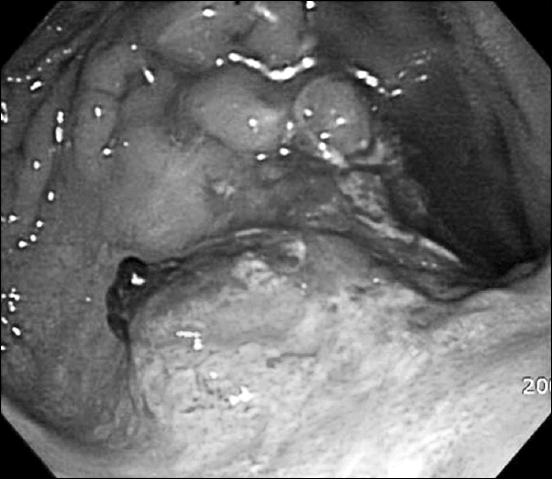
Fig. 1. Gastroscopic findings. A flat elevated mass was located on the posterior wall of upper body and fundus, measuring about 2 cm in size. It had an irregular surface with suspected area of vessel exposure.
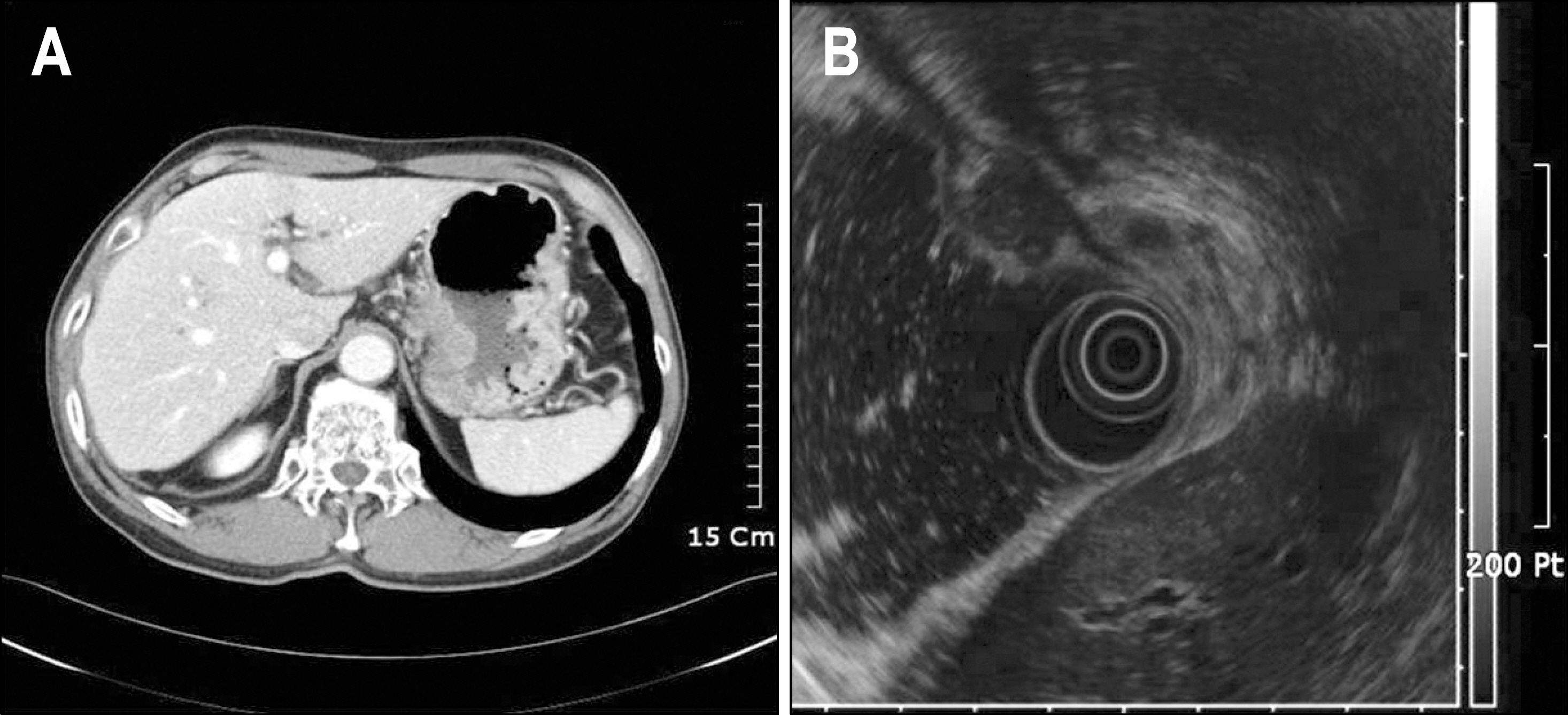
Fig. 2. Abdominal CT and EUS findings. (A) Abdominal CT showed focal irregular thickening of posterior wall of body and fundal area with small regional lymphadenopa-thies. (B) A 2 cm sized heteroge-nous, hypoechoic mass was seen on the posterior wall of upper body, involving proper muscle layer. Regional lymphadenopathy was suspicious.

Fig. 3. The gross specimen. (A) Two discrete nodular masses were located on the posterior wall of the gastric body and fundus, measuring 3×3 cm and 1×1 cm (arrow). (B) Cut surface showed grayish white solid masses involving up to muscular and submucosal layers for larger and smaller one, respectively.

Fig. 4. Microscopic findings. (A) The tumor was composed of large epithelioid cells without any organoid structure (H&E stain, ×40). (B) The tumor cells had abundant eosinophilic cytoplasm, well defined cell borders, and oval to irregular nuclei with vesicular chromatin and large eosinophilic nucleoli. Some tumor cells showed prominent clear or foamy cytoplasm. Mitotic figures were also seen (H&E stain, ×400).

Fig. 5. Immunohistochemical stain. Tumor cells were positive for (A) CD68 (×200), and (B) lysozyme (×200).
Reference
-
1. Hornick JL, Jaffe ES, Fletcher CD. Extranodal histiocytic sarcoma: clinicopathologic analysis of 14 cases of a rare epithelioid malignancy. Am J Surg Pathol. 2004; 28:1133–1144.2. Chan JKC. Tumors of the lymphoreticular system, including spleen and thymus. Fletcher CD, editor. Diagnostic histopathology of tumors. 2nd ed.London: Churchill Livingstone;2000. p. 1198–1199.3. Chun HJ, Kee KH, Suh CH, Lim SC, Song HS. Primary malignant lymphoma of true histiocytic origin of the liver: histiocytic sarcoma, kupffer cell sarcoma: a case report with immunohistochemical and ultrastructural studies. Korean J Pathol. 1989; 23:165–180.4. Kim KW, Park SY, Kim HJ, et al. A case with disseminated macrophage-related histiocytic sarcoma diagnosed by positive histiocytic markers. Korean J Hematol. 1999; 34:641–645.5. Jung SH, Jeong HJ, Jung WH, Kim TS, Choi IJ. Histopa-thological and immunocytochemical studies of primary gastrointestinal lymphomas in Korean patients. Korean J Pathol. 1987; 21:153–167.6. Zhang X, Kryston JJ, Michalak WA, Zhang K, Lin F, Schuerch C. Histiocytic sarcoma in the small intestine: a case report with flow cytometry study and review of the literature. Pathol Res Pract. 2008; 204:763–770.
Article7. Huang SC, Chang CL, Huang CH, Chang CC. Histiocytic sarcoma - a case with evenly distributed multinucleated giant cells. Pathol Res Pract. 2007; 203:683–689.
Article8. Favara BE, Feller AC, Pauli M, et al. Contemporary classification of histiocytic disorders. Med Pediatr Oncol. 1997; 29:157–166.
Article9. Paik JH, Jeon YK, Park SS, et al. Histiocytic sarcoma of the spleen: a case report and review of the literature. Korean J Pathol. 2005; 39:356–359.10. Yoshida C, Takeuchi M. Histiocytic sarcoma: identification of its histiocytic origin using immunohistochemistry. Intern Med. 2008; 47:165–169.
Article11. Vos JA, Abbondanzo SL, Barekman CL, Andriko JW, Miettinen M, Aguilera NS. Histiocytic sarcoma: a study of five cases including the histiocyte marker CD163. Mod Pathol. 2005; 18:693–704.
Article12. Kobayashi S, Kimura F, Hama Y, et al. Histiocytic sarcoma of the spleen: case report of asymptomatic onset of thrombo-cytopenia and complex imaging features. Int J Hematol. 2008; 87:83–87.
Article13. De Vos FY, Gerding MN, Arends JW, Wegman JJ. Histiocytic sarcoma localised in the thyroid: a case report. Ann Hematol. 2008; 87:681–682.14. Pileri SA, Grogan TM, Harris NL, et al. Tumours of histio-cytes and accessory dendritic cells: an immunohistochemical approach to classification from the International Lymphoma Study Group based on 61 cases. Histopathology. 2002; 41:1–29.
Article15. Audouin J, Vercelli-Retta J, Le Tourneau A, et al. Primary histiocytic sarcoma of the spleen associated with erythrophag-ocytic histiocytosis. Pathol Res Pract. 2003; 199:107–112.
Article16. Fukunaga M, Kato H. Histiocytic sarcoma associated with idi-opathic myelofibrosis. Arch Pathol Lab Med. 2004; 128:1167–1170.
Article17. Avilés-Salas A, Peñ a-Torres Mde L, Molina-Cruz A, Rivas- Vera S. Histiocytic sarcoma of the small intestine: report of one case. Rev Med Chil. 2009; 137:269–274.18. Abidi MH, Tove I, Ibrahim RB, Maria D, Peres E. Thalido-mide for the treatment of histiocytic sarcoma after hematopoietic stem cell transplant. Am J Hematol. 2007; 82:932–933.
Article19. Yaman E, Ozturk B, Erdem O, et al. Histiocytic sarcoma: PET-CT evaluation of a rare entity. Ann Nucl Med. 2008; 22:715–717.
Article
- Full Text Links
-
- Actions
-
Cited
- CITED
-
- Close
- Share
-
- Similar articles
-
- Collision Tumor Composed of a Granulocytic Sarcoma and an Adenocarcinoma of the Stomach: A Case Report
- Histiocytic Sarcoma of Rectum: A Case Report
- A Case of Carcinosarcoma and Adenocarcinoma of the Stomach
- Two cases of mucinous adenocarcinoma of the stomach mistaken as submucosal tumor
- Papillary gastric adenocarcinoma
