Korean J Gastroenterol.
2013 Jun;61(6):343-346. 10.4166/kjg.2013.61.6.343.
A Case of Primary Aortoenteric Fistula Mimicking Ulcer Bleeding
- Affiliations
-
- 1Department of Internal Medicine, Yuseong Sun Hospital, Daejeon, Korea.
- 2Department of Internal Medicine, Chungbuk National University College of Medicine, Cheongju, Korea. joungho@cbnu.ac.kr
- 3Department of Radiology, Chungbuk National University College of Medicine, Cheongju, Korea.
- KMID: 1718067
- DOI: http://doi.org/10.4166/kjg.2013.61.6.343
Abstract
- Primary aortoenteric fistula (PAEF) is a rare disease with a high mortality rate due to massive hemorrhaging and diagnostic difficulties. Although hemorrhagic regions can be identified by endoscopy, it is difficult to diagnose PAEF by this method. If PAEF is suspected, endoscopic procedure should be terminated and abdominal CT should be performed. Overlooking the herald bleeding of PAEF can lead to massive bleeding and death. An 85-year-old previously healthy male presented with a complaint of melena. Gastrointestinal endoscopy identified a hemorrhagic site in the third portion of the duodenum and endoscopic hemostasis was performed. However, during the procedure, it became apparent that the hemorrhage was probably not the result of a simple duodenal ulceration and abdominal CT was performed immediately. An aortic aneurysm connected to the duodenum was identified, confirming the diagnosis of PAEF. However, the patient died of massive hemorrhaging before an operation could be performed.
Keyword
MeSH Terms
Figure
-
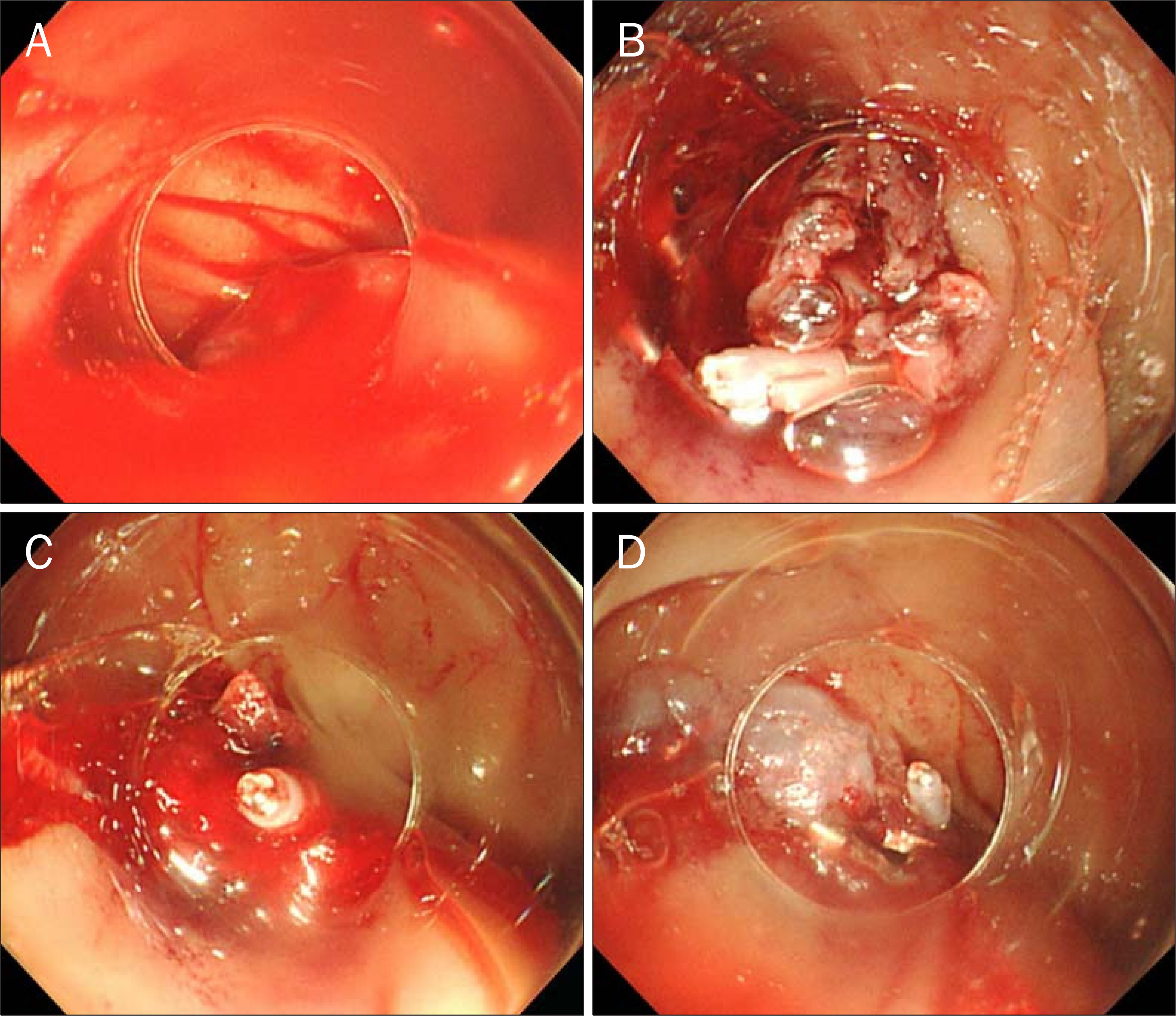
Fig. 1. Endoscopic findings. (A) Fresh duodenal bleeding after blood clot removal. (B, C) Continuous bleeding after clipping of the necrotized mucous membrane. (D) Stoppage of the bleeding after fibrin glue injection.
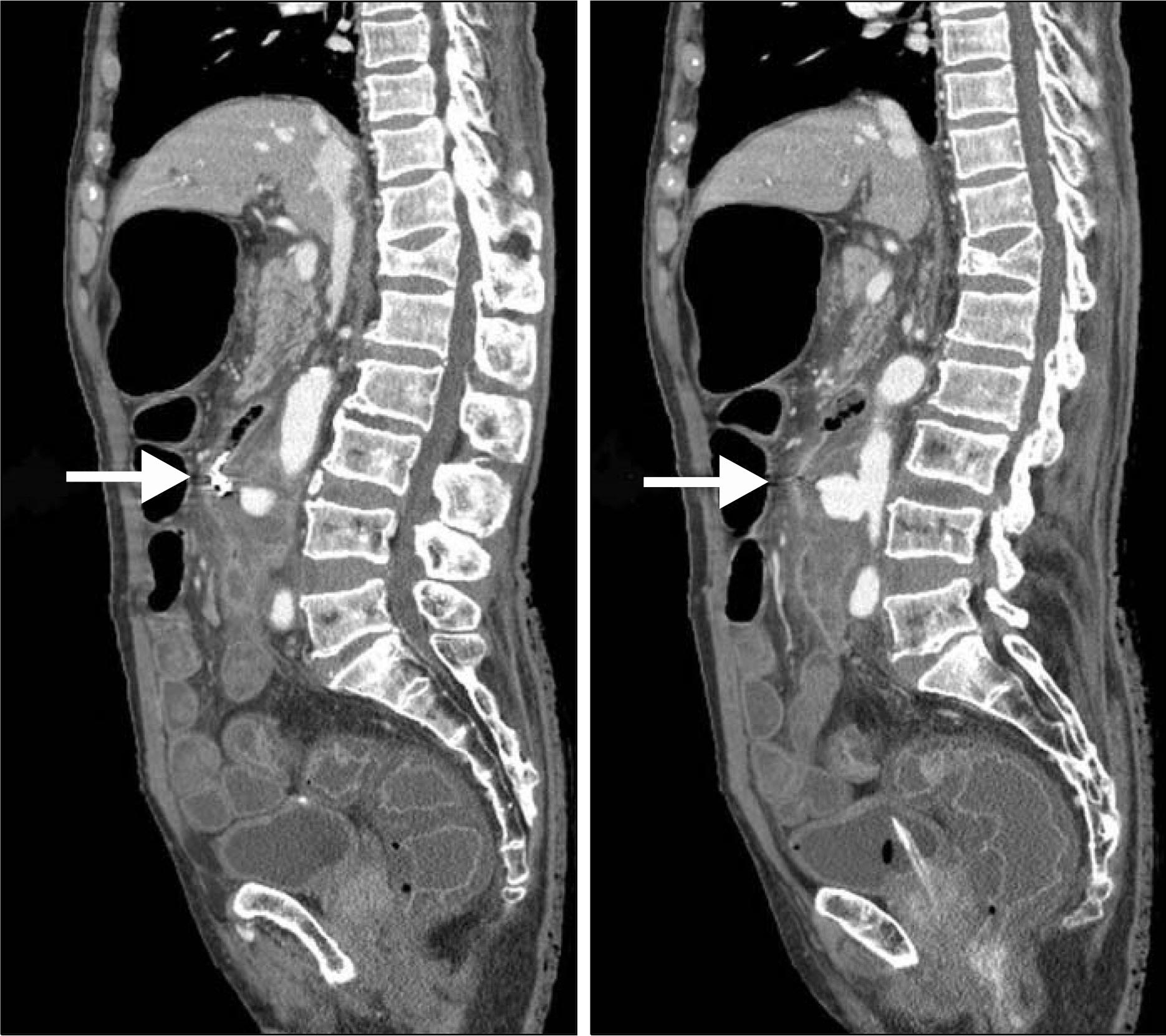
Fig. 2. Transverse abdominal CT showing communications between the abdominal aortic aneurysm (about 3.5 cm in diameter) and the third portion of the duodenum (visible hemo clips) (arrows).

Fig. 3. Sagittal abdominal CT showing communications between the saccular abdominal aortic aneurysm of the abdominal aorta (lumbar levels 3–4) and the distal duodenum (arrow).
Reference
-
References
1. Lemos DW, Raffetto JD, Moore TC, Menzoian JO. Primary aortoduodenal fistula: a case report and review of the literature. J Vasc Surg. 2003; 37:686–689.
Article2. Lee JT, Saroyan RM, Belzberg G, Pianim NA, Bongard FS. Primary aortoenteric fistula: computed tomographic diagnosis of an atypical presentation. Ann Vasc Surg. 2001; 15:251–254.
Article3. Saers SJ, Scheltinga MR. Primary aortoenteric fistula. Br J Surg. 2005; 92:143–152.
Article4. Ihama Y, Miyazaki T, Fuke C, et al. An autopsy case of a primary aortoenteric fistula: a pitfall of the endoscopic diagnosis. World J Gastroenterol. 2008; 14:4701–4704.
Article5. Lozano FS, Muñoz-Bellvis L, San Norberto E, Garcia-Plaza A, Gonzalez-Porras JR. Primary aortoduodenal fistula: new case reports and a review of the literature. J Gastrointest Surg. 2008; 12:1561–1565.
Article6. Song Y, Liu Q, Shen H, Jia X, Zhang H, Qiao L. Diagnosis and management of primary aortoenteric fistulas-experience learned from eighteen patients. Surgery. 2008; 143:43–50.
Article7. Tozzi FL, da Silva ES, Campos F, Fagundes Neto HO, Lucon M, Lupinacci RM. Primary aortoenteric fistula related to septic aortitis. Sao Paulo Med J. 2001; 119:150–153.
Article8. Baril DT, Carroccio A, Ellozy SH, et al. Evolving strategies for the treatment of aortoenteric fistulas. J Vasc Surg. 2006; 44:250–257.
Article9. Shree D, Jeppu S, Puneet P, Rani K. Computed tomography diagnosis of a primary aortoduodenal fistula in a patient with a partially thrombosed abdominal aortic aneurysm. Jpn J Radiol. 2010; 28:534–537.
Article10. Mylona S, Ntai S, Pomoni M, Kokkinaki A, Lepida N, Thanos L. Aortoenteric fistula: CT findings. Abdom Imaging. 2007; 32:393–397.
Article11. Sugai K, Kajiwara E, Mochizuki Y, et al. Intramural duodenal hematoma after endoscopic therapy for a bleeding duodenal ulcer in a patient with liver cirrhosis. Intern Med. 2005; 44:954–957.
Article12. Sadio A, Peixoto P, Cancela E, et al. Intramural hematoma: a rare complication of endoscopic injection therapy for bleeding peptic ulcers. Endoscopy. 2011; 43(Suppl 2 UCTN):E141–E142.
Article
- Full Text Links
-
- Actions
-
Cited
- CITED
-
- Close
- Share
-
- Similar articles
-
- Primary Aortoduodenal Fistula Causes Massive Melena: A Case Report
- A Case of Primary Aortoenteric Fistula Mimicking Duodenal Subepithelial Tumor
- Double Primary Aortoenteric Fistulae: A Case Report of Two Simultaneous Primary Aortoenteric Fistulae in One Patient
- A Rare but Fatal Instance of Gastrointestinal Bleeding: Primary Aortoenteric Fistula
- Primary Aortoenteric Fistula of a Saccular Aneurysm: Case Study and Literature Review
