Inflammatory Pseudotumor of the Liver Treated by Hepatic Resection: A Case Report
- Affiliations
-
- 1Department of Surgery, Yonsei University College of Medicine, Seoul, Korea. choi5491@yumc.yonsei.ac.kr
- 2Department of Pathology, Yonsei University College of Medicine, Seoul, Korea.
- KMID: 1715885
- DOI: http://doi.org/10.3349/ymj.2006.47.1.140
Abstract
- Inflammatory pseudotumor (IPT) of the liver is rare benign tumor. When the diagnosis of IPT is established with biopsy, simple observation or conservative therapy is preferred because of the possibility of regression. But IPT is unresponsive to the conservative treatment, surgical resection should be considered. We experienced a 63-year-old male, who was suspected hepatocellular carcinoma in abdominal computed tomography (CT) and magnetic resonance image (MRI) scan, presented with 2-month history of intermittent fever and weight loss. Percutaneous ultrasound guided core biopsy confirmed IPT of the liver. Non-steroidal anti-inflammatory drugs and antibiotics were administered for 8 and 4 weeks, respectively, but fever continued. So, extended right hepatectomy was performed for IPT of the liver and then fever subsided. The patient remains well during a follow-up period of 12 months.
Keyword
MeSH Terms
Figure
-
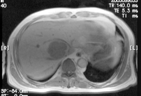
Fig. 1 Contrast enhanced T1 weighted MRI scan shows a relatively well-encapsulated, hypervascular mass, 6 cm in diameter on the right lobe of the liver.
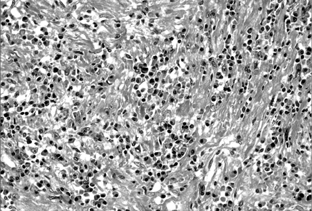
Fig. 2 Percutaneous biopsy shows the marked infiltration of lymphoplasma cells and some lymphocytes that are without cytologic atypia (H&E, × 400).

Fig. 3 Three months after the initial presentation, a follow-up contrast enhanced CT scan shows no interval changes of the tumor measuring 6 × 5.5 cm in Couinaud segment VIII of the liver.
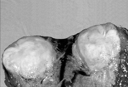
Fig. 4 The cut surface shows a yellowish-white nodule that is firm and well circumscribed.
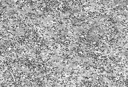
Fig. 5 Histologic findings of the tumor reveals a mixture of chronic inflammatory cells in which the polyclonal plasma cells predominate in a background of fibrotic stroma. (H&E, × 400).
Reference
-
1. Broughan TA, Fischer WL, Tuthill RJ. Vascular invasion by hepatic inflammatory pseudotumor. A clinicopathologic study. Cancer. 1993. 71:2934–2940.2. Byun YJ, Chung BH, Kwon KW. Inflammatory pseudotumor of urinary bladder. Yonsei Med J. 2000. 41:273–275.3. Choi SK, Choi YD, Cheon SH, Byun Y, Cho SW. Inflammatory pseudotumor of the urinary bladder in a child. Yonsei Med J. 2000. 41:401–403.4. Pack GT, Baker HW. Total right hepatic lobectomy: report of a case. Ann Surg. 1953. 138:253–258.5. Torzilli G, Inoue K, Midorikawa Y, Hui AM, Takayama T, Makuuchi M. Inflammatory pseudotumors of the liver: prevalence and clinical impact in surgical patients. Hepatogastroenterology. 2001. 48:1118–1123.6. Jais P, Berger JF, Vissuzaine C, Paramelle O, Clays-Schouman E, Potet F, et al. Regression of inflammatory pseudotumor of the liver under conservative therapy. Dig Dis Sci. 1995. 40:752–756.7. White JE, Chase CW, Kelley JE, Brock WB, Clark MO. Inflammatory pseudotumor of the liver associated with extrahepatic infection. South Med J. 1997. 90:23–29.8. Nam KJ, Kang HK, Lim JH. Inflammatory pseudotumor of the liver: CT and sonographic findings. AJR. 1996. 167:485–487.9. Schwarz RE, Reynolds JC, Lotze MT. "Pseudo-pseudotumor" of the liver. Adenocarcinoma presenting as an inflammatory pseudotumor. Dig Dis Sci. 1994. 39:2679–2684.10. Koea JB, Broadhurst GW, Rodgers MS, McCall JL. Inflammatory pseudotumor of the liver: demographics, diagnosis, and the case for nonoperative management. J Am Coll Surg. 2003. 196:226–235.11. Gluszek S, Kot M, Czerwaty M. Inflammatory pseudotumor of the liver treated surgically. Hepatogastroenterology. 1999. 46:2959–2960.12. Lu CC, Chen CC, Hsia CY, Chiang JH, Tsay SH, Han HF, et al. A progressive growing inflammatory pseudotumor of the liver. Zhonghua Yi Xue Za Zhi (Taipei). 2001. 64:725–730.13. Kaneko K, Ando H, Watanabe Y, Seo T, Nagino M, Kamiya J, et al. Aggressive preoperative management and extended surgery for inflammatory pseudotumor involving the hepatic hilum in a child. Surgery. 2001. 129:757–760.14. Pecorella I, Ciardi A, Memeo L, Trombetta G, de Quarto A, de Simone P, et al. Inflammatory pseudotumour of the liver-evidence for malignant transformation. Pathol Res Pract. 1999. 195:115–120.15. Zavaglia C, Barberis M, Gelosa F, Cimino G, Minola E, Mondazzi L, et al. Inflammatory pseudotumour of the liver with malignant transformation. Report of two cases. Ital J Gastroenterol. 1996. 28:152–159.
- Full Text Links
-
- Actions
-
Cited
- CITED
-
- Close
- Share
-
- Similar articles
-
- Spontaneous Regression of Inflarmmatory Pseudotumor of the Liver by Conservative Therapy
- Inflammatory Pseudotumor of the Liver
- Inflammatory Pseud0tumor of the Liver: A case report
- Inflammatory Pseudotumor in the Liver and Right Omentum Caused by Pelvic Inflammatory Disease: A Case Report
- Inflammatory Pseudotumor of the Liver: A case report
