Anterior Commissure - Posterior Commissure Revisited
- Affiliations
-
- 1Neuroscience Research Institute, Gachon University of Medicine and Science, Incheon 405-760, Korea. zcho@gachon.ac.kr
- KMID: 1715771
- DOI: http://doi.org/10.3348/kjr.2013.14.4.653
Abstract
OBJECTIVE
The anterior commissure (AC) and posterior commissure (PC) are the two distinct anatomic structures in the brain which are difficult to observe in detail with conventional MRI, such as a 1.5T MRI system. However, recent advances in ultra-high resolution MRI have enabled us to examine the AC and PC directly. The objective of the present study is to standardize the shape and size of the AC and PC using a 7.0T MRI and to propose a new brain reference line.
MATERIALS AND METHODS
Thirty-four, 21 males and 13 females, healthy volunteers were enrolled in this study. After determining the center of each AC and PC, we defined the connection of these centers as the central intercommissural line (CIL). We compared the known extra- and intra-cerebral reference lines with the CIL to determine the difference in the angles. Additionally, we obtained horizontal line from flat ground line of look front human.
RESULTS
The difference in angle of the CIL and the tangential intercommissural line (TIL) from the horizontal line was 8.7 +/- 5.1 (11 +/- 4.8) and 17.4 +/- 5.2 (19.8 +/- 4.8) degrees in males and females, respectively. The difference in angle between the CIL and canthomeatal line was 10.1 in both male and female, and there was no difference between both sexes. Likewise, there was no significant difference in angle between the CIL and TIL between both sexes (8.3 +/- 1.1 in male and 8.8 +/- 0.7 in female).
CONCLUSION
In this study, we have used 7.0T MRI to define the AC and PC quantitatively and in a more robust manner. We have showed that the CIL is a reproducible reference line and serves as a standard for the axial images of the human brain.
Keyword
MeSH Terms
Figure
-
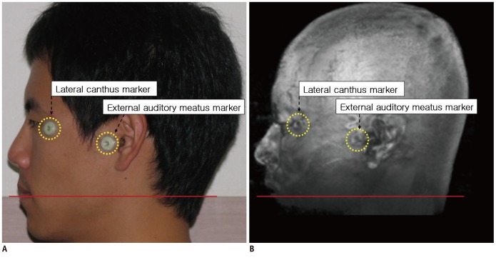
Fig. 1 Photograph and MR-rendered images of single volunteer.A. Photograph of volunteer. B. Three-dimensional volume rendering low resolution magnetization prepared rapid gradient echo image. Yellow circles indicate markers. Red line indicates horizontal line.
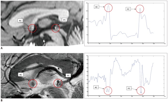
Fig. 2 Comparison of AC and PC in 1.5T and 7.0T images (sagittal view).A. 1.5T image with central commissural line (black dotted line) and intensity plot (blue solid line). B. Same as (A) with 7.0T image. AC and PC are circled in sagittal views of both 1.5T and 7.0T MRI images. Note clear AC and PC contrast and edge in 7.0T image. AC = anterior commissure, PC = posterior commissure
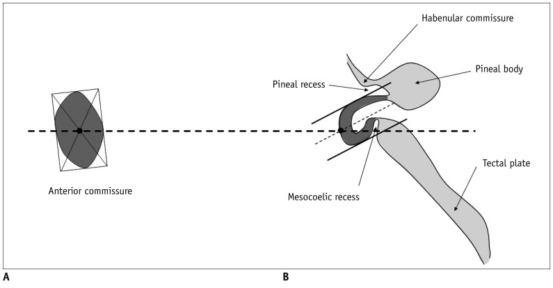
Fig. 3 Quantification of AC and PC.A. Method of defining center point in AC. B. Method of determining center point in PC in reference to pineal and mesocoelic recesses. In PC, we have selected midpoint of C-shape arch of PC. AC = anterior commissure, PC = posterior commissure
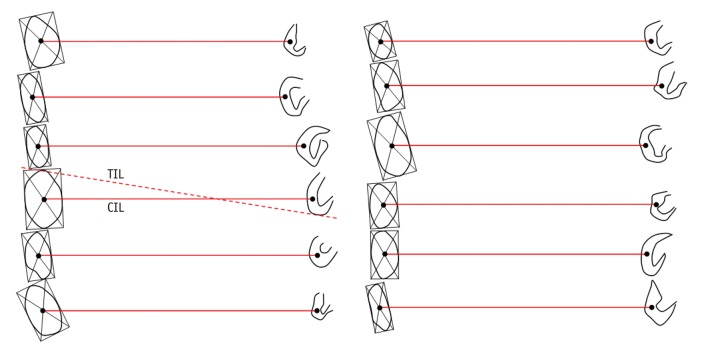
Fig. 4 Examples of center points of actual AC and PC, which make CIL we have defined.Tangential intercommissural line (TIL) is also shown for reference in one example. AC = anterior commissure, PC = posterior commissure, CIL = central intercommissural line, TIL = tangential intercommissural line
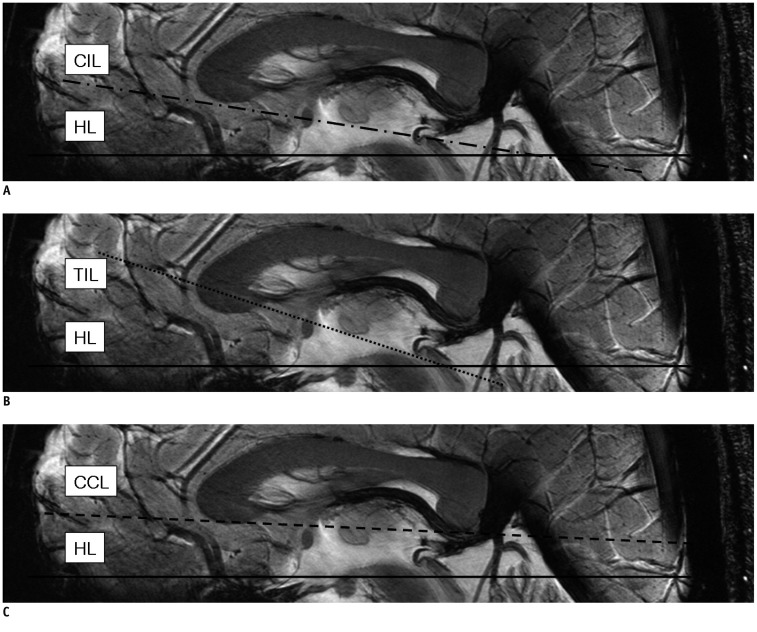
Fig. 5 Various intercommissural and extended cerebral reference lines in reference to horizontal line (HL : solid line).A. Central intercommissural line (CIL : dash-dot line). B. Tangential intercommissural line (TIL : dotted line). C. Corpus callosum line (CCL : dashed line). CCL = corpus callosum line, CIL = central intercommissural line, HL = horizontal line, TIL = tangential intercommissural line
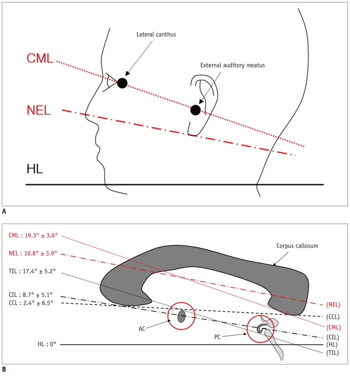
Fig. 6 Extracerebral & intracerebral reference lines in reference to horizontal line.A. Extracerebral reference lines : nose-ear line (NEL : red dash-dot line), canthomeatal line (CML : red dotted line) all in reference to horizontal line. B. Summary of result in case of male. AC = anterior commissure, PC = posterior commissure, CCL = corpus callosum line (black dashed line), CIL = central intercommissural line (black dash-dot line), CML = canthomeatal line (red dotted line), HL = horizontal line (black solid line), NEL = nose-ear line (red dash-dot line), TIL = tangential intercommissural line (black dotted line)
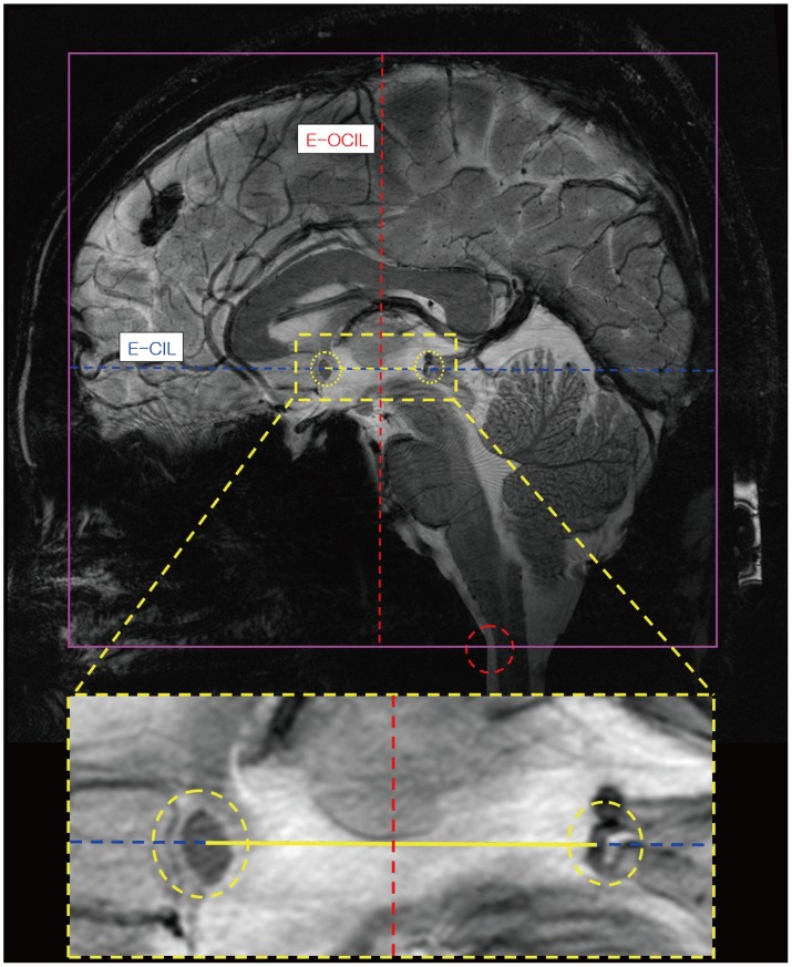
Fig. 7 Definition of virtual extension lines: Blue dotted line indicates extended central lintercommussural line.Yellow line at middle indicates anterior commissure-posterior commissure connecting line and red dotted line is extended orthogonal central intercommissural line. Two yellow circles show AC and PC points. Red circle is reference point of inferior brain part. AC = anterior commissure, PC = posterior commissure
Reference
-
1. Cho ZH, Chung MS, Chi JG, Na DL. 7.0 Tesla MRI Brain Atlas: In Vivo Atlas with Cryomacrotome Correlation. Heidelberg: Springer;2010.2. Cho ZH, Kim YB, Han JY, Min HK, Kim KN, Choi SH, et al. New brain atlas-mapping the human brain in vivo with 7.0 T MRI and comparison with postmortem histology: will these images change modern medicine? Int J Imaging Syst Technol. 2008; 18:2–8.
Article3. Weiss KL, Pan H, Storrs J, Strub W, Weiss JL, Jia L, et al. Clinical brain MR imaging prescriptions in Talairach space: technologist- and computer-driven methods. AJNR Am J Neuroradiol. 2003; 24:922–929. PMID: 12748095.4. Nowinski WL. Modified Talairach landmarks. Acta Neurochir (Wien). 2001; 143:1045–1057. PMID: 11685613.
Article5. Choi DY, Chung SC, Lee BY, Eom JS, Sohn JH. Development of Korean standard brain templates according to gender and age. Korean J Anat. 2004; 37:255–261.6. Talairach J, Tournoux P. Co-planar Stereotaxic Atlas of the Human Brain. New York: Thieme;1988.7. Schaltenbrand G, Wahren W, Hassler RG. Atlas of Stereotaxy of the Human Brain. 2nd ed. Thieme: Stuttgart;1977.8. Lee U, Kim YB, Kim MH, Ahn SG, Kang DS, Park CW. Basic estimation of intracerebral reference points: data analysis from 169 Korean people. J Korean Neurosurg Soc. 1995; 24:1056–1060.
- Full Text Links
-
- Actions
-
Cited
- CITED
-
- Close
- Share
-
- Similar articles
-
- The Normal Width of the Anterior Commissure of the True Vocal Cord in Korean Adults Measured by Helical CT
- Avulsion of Aortic Commissure: Rare Cause of Aortic Regurgitation: 2 case reports
- A Rare Midbrain Syndrome with Bilateral Cerebellar Ataxia and Isolated Superior Rectus Palsy: Wernekinck Commissure Syndrome
- Interthalamic adhesion in humans: a gray commissure?
- A New 'Y' Shape Partial Laryngectomy for Supraglottic Carcinoma with Anterior Commissure Invasion or Encroachment
